2F0X

Crystal structure and function of human thioesterase superfamily member 2(THEM2)


Experimental Data Snapshot

  • Method: X-RAY DIFFRACTION
  • Resolution: 2.30 Å
  • R-Value Free: 0.247 
  • R-Value Work: 0.216 
  • R-Value Observed: 0.219 

wwPDB Validation   3D Report Full Report


This is version 1.3 of the entry. See complete history


Literature

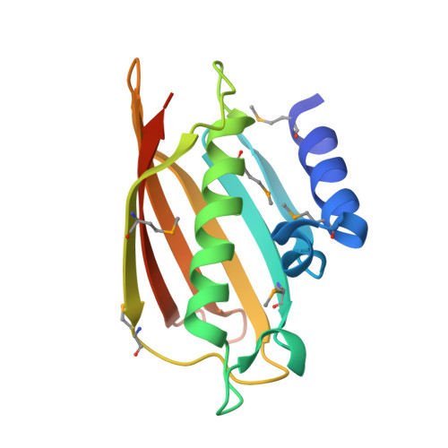
Crystal structure of human thioesterase superfamily member 2

Cheng, Z.Song, F.Shan, X.Wei, Z.Wang, Y.Dunaway-Mariano, D.Gong, W.

(2006) Biochem Biophys Res Commun 349: 172-177

  • DOI: https://doi.org/10.1016/j.bbrc.2006.08.025
  • Primary Citation of Related Structures:  
    2F0X

  • PubMed Abstract: 

    Hotdog-fold has been identified in more than 1000 proteins, yet many of which in eukaryotes are less studied. No structural or functional studies of human thioesterase superfamily member 2 (hTHEM2) have been reported before. Since hTHEM2 exhibits about 20% sequence identity to Escherichia coli PaaI protein, it was proposed to be a thioesterase with a hotdog-fold. Here, we report the crystallographic structure of recombinant hTHEM2, determined by the single-wavelength anomalous dispersion method at 2.3A resolution. This structure demonstrates that hTHEM2 indeed contains a hotdog-fold and forms a back-to-back tetramer as other hotdog proteins. Based on structural and sequence conservation, the thioesterase active site in hTHEM2 is predicted. The structure and substrate specificity are most similar to those of the bacterial phenylacetyl-CoA hydrolase. Asp65, located on the central alpha-helix of subunit B, was shown by site-directed mutagenesis to be essential to catalysis.


  • Organizational Affiliation

    School of Life Sciences, University of Science and Technology of China, Hefei, Anhui 230026, PR China.


Macromolecules
Find similar proteins by:  (by identity cutoff)  |  3D Structure
Entity ID: 1
MoleculeChains Sequence LengthOrganismDetailsImage
Thioesterase superfamily member 2
A, B, C, D, E
A, B, C, D, E, F, G, H
148Homo sapiensMutation(s): 7 
UniProt & NIH Common Fund Data Resources
Find proteins for Q9NPJ3 (Homo sapiens)
Explore Q9NPJ3 
Go to UniProtKB:  Q9NPJ3
PHAROS:  Q9NPJ3
GTEx:  ENSG00000112304 
Entity Groups  
Sequence Clusters30% Identity50% Identity70% Identity90% Identity95% Identity100% Identity
UniProt GroupQ9NPJ3
Sequence Annotations
Expand
  • Reference Sequence
Small Molecules
Ligands 1 Unique
IDChains Name / Formula / InChI Key2D Diagram3D Interactions
SO4
Query on SO4

Download Ideal Coordinates CCD File 
AA [auth H]
BA [auth H]
I [auth A]
J [auth A]
K [auth B]
AA [auth H],
BA [auth H],
I [auth A],
J [auth A],
K [auth B],
L [auth B],
M [auth B],
N [auth C],
O [auth C],
P [auth C],
Q [auth C],
R [auth D],
S [auth D],
T [auth E],
U [auth E],
V [auth E],
W [auth F],
X [auth F],
Y [auth G],
Z [auth G]
SULFATE ION
O4 S
QAOWNCQODCNURD-UHFFFAOYSA-L
Modified Residues  1 Unique
IDChains TypeFormula2D DiagramParent
MSE
Query on MSE
A, B, C, D, E
A, B, C, D, E, F, G, H
L-PEPTIDE LINKINGC5 H11 N O2 SeMET
Experimental Data & Validation

Experimental Data

  • Method: X-RAY DIFFRACTION
  • Resolution: 2.30 Å
  • R-Value Free: 0.247 
  • R-Value Work: 0.216 
  • R-Value Observed: 0.219 
  • Space Group: C 1 2 1
Unit Cell:
Length ( Å )Angle ( ˚ )
a = 131.937α = 90
b = 122.076β = 123.9
c = 90.977γ = 90
Software Package:
Software NamePurpose
DENZOdata reduction
SCALEPACKdata scaling
SHELXphasing
RESOLVEphasing
CNSrefinement
PDB_EXTRACTdata extraction
HKL-2000data reduction
SHELXDphasing

Structure Validation

View Full Validation Report


Entry History 

Deposition Data

Revision History  (Full details and data files)

  • Version 1.0: 2006-10-10
    Type: Initial release
  • Version 1.1: 2008-05-01
    Changes: Version format compliance
  • Version 1.2: 2011-07-13
    Changes: Version format compliance
  • Version 1.3: 2017-10-18
    Changes: Refinement description