Structures of oligosaccharide-bound forms of the endoglucanase V from Humicola insolens at 1.9 A resolution.
Davies, G.J., Tolley, S.P., Henrissat, B., Hjort, C., Schulein, M.(1995) Biochemistry 34: 16210-16220
- PubMed: 8519779 Search on PubMed
- DOI: https://doi.org/10.1021/bi00049a037
- Primary Citation Related Structures:
2ENG - PubMed Abstract:
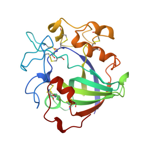
Cellulose, a polymer of beta-1,4-linked glucose residues, is the major polysaccharide component of plant cell walls and the most abundant biopolymer. The underlying mechanisms of the enzymatic degradation of cellulose are of increasing commercial and ecological significance. Endoglucanase V, from the cellulolytic soil hyphomycete Humicola insolens, is an endocellulase, the catalytic core of which consists of 210 amino acids and is known to hydrolyze the beta-1,4 links with inversion of configuration at the anomeric carbon. The major products of cellulose hydrolysis are cellobiose and cellotriose. The crystal structures of the endoglucanase V (EGV) from H. insolens, in native, product (cellobiose), inactive mutant (D10N), and oligosaccharide-bound [(D10N)-cellohexaose] forms, have been determined at resolutions of 1.9 A or better. EGV consists of a six-stranded beta-barrel domain with long interconnecting loops. A 40 A groove exists along the surface of the enzyme, and this contains the catalytic residues, Asp 10 and Asp 121. The two catalytic aspartates sit to either side of the substrate binding groove in an ideal conformation for facilitating cleavage by inversion, their carboxyl groups being separated by approximately 8.5 A. The complex between substrate and inactive mutant reveals excellent density for an oligosaccharide in six of the enzyme's seven substrate binding subsites. No sugar moiety, however, is seen bound to the -1 subsite at the point of cleavage. The geometry of the cleavage site suggests that the enzyme would favor the binding of sugars with an elongated glycosidic bond, as found in the transition state, as opposed to the binding of substrate. The oligosaccharide complexes reveal solvent water suitably placed for participation in a single displacement reaction as first suggested by Koshland in 1953 [Koshland, D. E. (1953) Biol. Rev. 28, 416-436]. A large conformational change takes place upon substrate binding. This "lid flipping" has the effect of increasing the hydrophobic environment of the catalytic proton donor, enclosing the active site at the point of cleavage, and bringing a third aspartate (Asp 114) in close proximity to the substrate. Site-directed mutagenesis of the catalytic residues has been used to confirm their significance in catalysis.
- Department of Chemistry, University of York, Heslington, England.
Organizational Affiliation: