Structural Polymorphism of Chromodomains in Chd1
Okuda, M., Horikoshi, M., Nishimura, Y.(2007) J Mol Biol 365: 1047-1062
- PubMed: 17098252 Search on PubMed
- DOI: https://doi.org/10.1016/j.jmb.2006.10.039
- Primary Citation Related Structures:
2DY7, 2DY8 - PubMed Abstract:
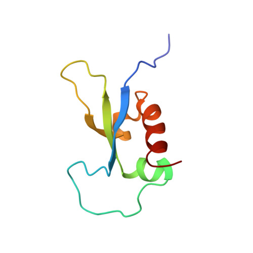
Chromodomain from heterochromatin protein 1 and polycomb protein is known to be a lysine-methylated histone H3 tail-binding module. Chromo-helicase/ATPase DNA-binding protein 1 (CHD1) is an ATP-dependent chromatin remodeling factor, containing two tandem chromodomains. In human CHD1, both chromodomains are essential for specific binding to a K4 methylated histone H3 (H3 MeK4) peptide and are found to bind cooperatively in the crystal structure. For the budding yeast homologue, Chd1, the second but not the first chromodomain was once reported to bind to an H3 MeK4 peptide. Here, we reveal that neither the second chromodomain nor a region containing tandem chromodomains from yeast Chd1 bind to any lysine-methylated or arginine-methylated histone peptides that we examined. In addition, we examined the structures of the chromodomains from Chd1 by NMR. Although the tertiary structure of the region containing tandem chromodomains could not be obtained, the secondary structure deduced from NMR is well conserved in the tertiary structures of the corresponding first and second chromodomains determined individually by NMR. Both chromodomains of Chd1 demonstrate a structure similar to that of the corresponding part of CHD1, consisting of a three-stranded beta-sheet followed by a C-terminal alpha-helix. However, an additional helix between the first and second beta-strands, which is found in both of the first chromodomains of Chd1 and CHD1, is positioned in an entirely different manner in Chd1 and CHD1. In human CHD1 this helix forms the peptide-binding site. The amino acid sequences of the chromodomains could be well aligned on the basis of these structures. The alignment showed that yeast Chd1 lacks several key functional residues, which are responsible for specific binding to a methylated lysine residue in other chromodomains. Chd1 is likely to have no binding affinity for any H3 MeK peptide, as found in other chromodomain proteins.
- Graduate School of Supramolecular Biology, Yokohama City University, Tsurumi-ku, Yokohama 230-0045, Japan.
Organizational Affiliation: