


Crystal Structure of Alanine Racemase from Corynebacterium glutamicum at 2.1 A resolution
Miyaguchi, I., Sasaki, C., Kato, R., Oikawa, T., Sugio, S.To be published.
Experimental Data Snapshot
Starting Model: experimental
View more details
Entity ID: 1 | |||||
|---|---|---|---|---|---|
| Molecule | Chains | Sequence Length | Organism | Details | Image |
| Alanine racemase | 361 | Corynebacterium glutamicum | Mutation(s): 0 Gene Names: alr EC: 5.1.1.1 | 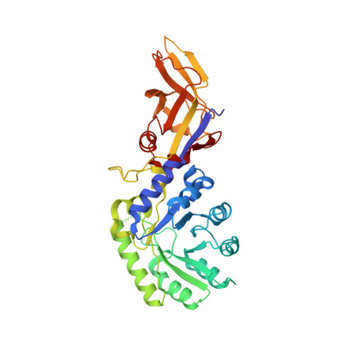 | |
UniProt | |||||
Entity Groups | |||||
| Sequence Clusters | 30% Identity50% Identity70% Identity90% Identity95% Identity100% Identity | ||||
| UniProt Group | Q8RSU9 | ||||
Sequence AnnotationsExpand | |||||
Reference Sequence | |||||
| Ligands 1 Unique | |||||
|---|---|---|---|---|---|
| ID | Chains | Name / Formula / InChI Key | 2D Diagram | 3D Interactions | |
| PLP Download:Ideal Coordinates CCD File | E [auth A], F [auth B], G [auth C], H [auth D] | PYRIDOXAL-5'-PHOSPHATE C8 H10 N O6 P NGVDGCNFYWLIFO-UHFFFAOYSA-N |  | ||
| Length ( Å ) | Angle ( ˚ ) |
|---|---|
| a = 78.384 | α = 90 |
| b = 113.612 | β = 94.73 |
| c = 88.096 | γ = 90 |
| Software Name | Purpose |
|---|---|
| HKL-2000 | data collection |
| CNS | refinement |
| HKL-2000 | data reduction |
| HKL-2000 | data scaling |
| CNS | phasing |


