2DMH | pdb_00002dmh

Solution structure of the first C2 domain of human myoferlin


Experimental Data Snapshot

  • Method: SOLUTION NMR
  • Conformers Calculated: 100 
  • Conformers Submitted: 20 
  • Selection Criteria: target function, structures with the lowest energy 

wwPDB Validation 3D Report Full Report

Validation slider image for 2DMH

This is version 1.4 of the entry. See complete history

Literature

Solution structure of the first C2 domain of human myoferlin

Nagashima, T.Hayashi, F.Yokoyama, S.

To be published.

Macromolecule Content 

  • Total Structure Weight: 15.02 kDa 
  • Atom Count: 1,056 
  • Modeled Residue Count: 140 
  • Deposited Residue Count: 140 
  • Unique protein chains: 1

Macromolecules

Find similar proteins by:|  3D Structure
Entity ID: 1
MoleculeChains  Sequence LengthOrganismDetailsImage
Myoferlin140Homo sapiensMutation(s): 0 
Gene Names: FER1L3KIAA1207MYOF
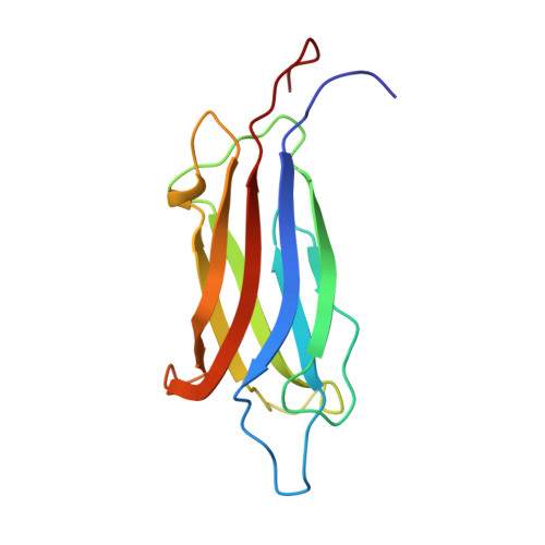
UniProt & NIH Common Fund Data Resources
Find proteins for Q9NZM1 (Homo sapiens)
Explore Q9NZM1 
Go to UniProtKB:  Q9NZM1
PHAROS:  Q9NZM1
GTEx:  ENSG00000138119 
Entity Groups
Sequence Clusters30% Identity50% Identity70% Identity90% Identity95% Identity100% Identity
UniProt GroupQ9NZM1
Sequence Annotations
Expand
Reference Sequence

Experimental Data & Validation

Experimental Data

  • Method: SOLUTION NMR
  • Conformers Calculated: 100 
  • Conformers Submitted: 20 
  • Selection Criteria: target function, structures with the lowest energy 

Structure Validation

View Full Validation Report


Entry History 

Deposition Data

Revision History  (Full details and data files)

  • Version 1.0: 2006-10-21
    Type: Initial release
  • Version 1.1: 2008-04-30
    Changes: Version format compliance
  • Version 1.2: 2011-07-13
    Changes: Version format compliance
  • Version 1.3: 2022-03-09
    Changes: Data collection, Database references, Derived calculations
  • Version 1.4: 2024-05-29
    Changes: Data collection