2DL1 | pdb_00002dl1

Solution structure of the MIT domain from human Spartin


Experimental Data Snapshot

  • Method: SOLUTION NMR
  • Conformers Calculated: 100 
  • Conformers Submitted: 20 
  • Selection Criteria: structures with the least restraint violation, target function 

wwPDB Validation 3D Report Full Report

Validation slider image for 2DL1

This is version 1.4 of the entry. See complete history

Literature

Solution structure of the MIT domain from human Spartin

Suetake, T.Hayashi, F.Yokoyama, S.

To be published.

Macromolecule Content 

  • Total Structure Weight: 12.77 kDa 
  • Atom Count: 895 
  • Modeled Residue Count: 116 
  • Deposited Residue Count: 116 
  • Unique protein chains: 1

Macromolecules

Find similar proteins by:|  3D Structure
Entity ID: 1
MoleculeChains  Sequence LengthOrganismDetailsImage
Spartin116Homo sapiensMutation(s): 0 
Gene Names: SPG20KIAA0610TAHCCP1
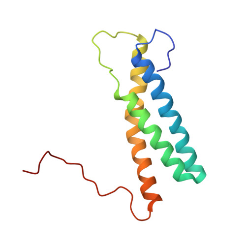
UniProt & NIH Common Fund Data Resources
Find proteins for Q8N0X7 (Homo sapiens)
Explore Q8N0X7 
Go to UniProtKB:  Q8N0X7
PHAROS:  Q8N0X7
GTEx:  ENSG00000133104 
Entity Groups
Sequence Clusters30% Identity50% Identity70% Identity90% Identity95% Identity100% Identity
UniProt GroupQ8N0X7
Sequence Annotations
Expand
Reference Sequence

Experimental Data & Validation

Experimental Data

  • Method: SOLUTION NMR
  • Conformers Calculated: 100 
  • Conformers Submitted: 20 
  • Selection Criteria: structures with the least restraint violation, target function 

Structure Validation

View Full Validation Report


Entry History 

Deposition Data

Revision History  (Full details and data files)

  • Version 1.0: 2006-10-14
    Type: Initial release
  • Version 1.1: 2008-04-30
    Changes: Version format compliance
  • Version 1.2: 2011-07-13
    Changes: Version format compliance
  • Version 1.3: 2022-03-09
    Changes: Data collection, Database references, Derived calculations
  • Version 1.4: 2024-05-29
    Changes: Data collection