2DI2 | pdb_00002di2

NMR structure of the HIV-2 nucleocapsid protein


Experimental Data Snapshot

  • Method: SOLUTION NMR
  • Conformers Calculated: 100 
  • Conformers Submitted: 13 
  • Selection Criteria: structures with the lowest energy, structures with the least restraint violations 

wwPDB Validation 3D Report Full Report

Validation slider image for 2DI2

This is version 1.5 of the entry. See complete history

Literature

RNA Recognition Mechanism of the Minimal Active Domain of the Human Immunodeficiency Virus Type-2 Nucleocapsid Protein

Matsui, T.Kodera, Y.Endoh, H.Miyauchi, E.Komatsu, H.Sato, K.Tanaka, T.Kohno, T.Maeda, T.

(2007) J Biochem 141: 269-277

  • DOI: https://doi.org/10.1093/jb/mvm037
  • Primary Citation Related Structures: 
    2DI2

  • PubMed Abstract: 

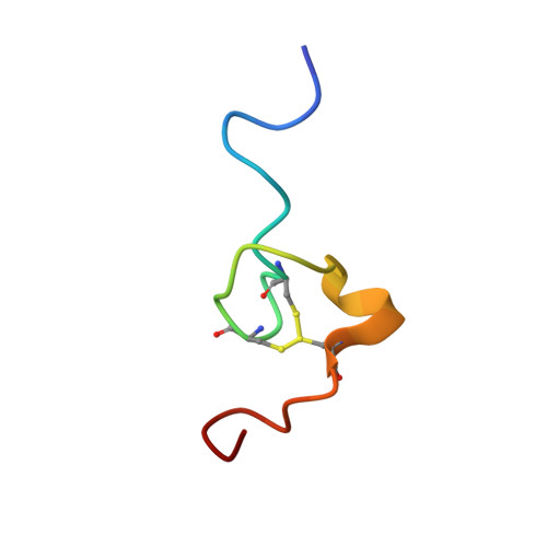
    NCp8 of HIV-2 contains two CCHC-type zinc fingers connected by a linker, and is involved in many critical steps of the virus life cycle. It was previously shown that the first zinc finger flanked by the linker is the minimal active domain for specific binding to viral RNA. In our previous study, we determined the three-dimensional structure of NCp8-f1, including the minimal active domain, and found that a hydrogen bond between Asn(11) N(delta)H and Arg(27) O stabilized the conformation of the linker in the vicinity of the zinc finger [Kodera et al. (1998) Biochemistry 37, 17704-17713]. In this study, RNA binding activities of NCp8-f1 and three types of its mutant peptides were analysed by native PAGE assay. The activity and three-dimensional structure of NCp8-f1/N11A, in which alanine is substituted for Asn(11) thereby affecting the conformation of the linker, was analyzed and compared with those of NCp8-f1. We demonstrated that the existence of Arg(4) and/or Lys(5) and Arg(26) and/or Arg(27) were necessary for binding RNA. Furthermore, the linker's flexible orientation, which is controlled by the hydrogen bond between Asn(11) N(delta)H and Arg(27) O, appears to be a structural basis for NCp8 existing as a multi-functional protein.


  • Organizational Affiliation
    • Department of Physics, School of Science, Kitasato University, Sagamihara, Kanagawa 228-8555, Japan.

Macromolecule Content 

  • Total Structure Weight: 3.41 kDa 
  • Atom Count: 233 
  • Modeled Residue Count: 29 
  • Deposited Residue Count: 29 
  • Unique protein chains: 1

Macromolecules

Find similar proteins by:|  3D Structure
Entity ID: 1
MoleculeChains  Sequence LengthOrganismDetailsImage
Nucleocapsid protein p729synthetic constructMutation(s): 1 
UniProt
Find proteins for P18041 (Human immunodeficiency virus type 2 subtype A (isolate Ghana-1))
Explore P18041 
Go to UniProtKB:  P18041
Entity Groups
Sequence Clusters30% Identity50% Identity70% Identity90% Identity95% Identity100% Identity
UniProt GroupP18041
Sequence Annotations
Expand
Reference Sequence

Small Molecules

Ligands 1 Unique
IDChains Name / Formula / InChI Key2D Diagram3D Interactions
ZN

Query on ZN


Download:Ideal Coordinates CCD File
B [auth A]ZINC ION
Zn
PTFCDOFLOPIGGS-UHFFFAOYSA-N

Experimental Data & Validation

Experimental Data

  • Method: SOLUTION NMR
  • Conformers Calculated: 100 
  • Conformers Submitted: 13 
  • Selection Criteria: structures with the lowest energy, structures with the least restraint violations 

Structure Validation

View Full Validation Report


Entry History 

Deposition Data

Revision History  (Full details and data files)

  • Version 1.0: 2007-03-13
    Type: Initial release
  • Version 1.1: 2008-04-30
    Changes: Version format compliance
  • Version 1.2: 2011-07-13
    Changes: Version format compliance
  • Version 1.3: 2019-12-11
    Changes: Advisory, Derived calculations, Source and taxonomy
  • Version 1.4: 2021-11-10
    Changes: Database references, Derived calculations
  • Version 1.5: 2024-05-29
    Changes: Data collection