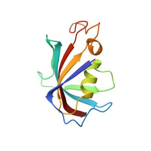
2DG4

FK506-binding protein mutant WF59 complexed with Rapamycin


Experimental Data Snapshot

  • Method: X-RAY DIFFRACTION
  • Resolution: 1.70 Å
  • R-Value Free: 0.213 
  • R-Value Work: 0.195 
  • R-Value Observed: 0.196 

wwPDB Validation   3D Report Full Report


Ligand Structure Quality Assessment 


This is version 1.4 of the entry. See complete history


Literature

Energetic and structural analysis of the role of tryptophan 59 in FKBP12

Fulton, K.F.Jackson, S.E.Buckle, A.M.

(2003) Biochemistry 42: 2364-2372

  • DOI: https://doi.org/10.1021/bi020564a
  • Primary Citation of Related Structures:  
    2DG3, 2DG4, 2DG9

  • PubMed Abstract: 

    Tryptophan 59 forms the seat of the hydrophobic ligand-binding site in the small immunophilin FKBP12. Mutating this residue to phenylalanine or leucine stabilizes the protein by 2.72 and 2.35 kcal mol(-1), respectively. Here we report the stability data and 1.7 A resolution crystal structures of both mutant proteins, complexed with the immunosuppressant rapamycin. Both structures show a relatively large response to mutation involving a helical bulge at the mutation site and the loss of a hydrogen bond that anchors a nearby loop. The increased stability of the mutants is probably due to a combination of improved packing and an entropic gain at the mutation site. The structures are almost identical to that of wild-type FKBP12.6, an isoform of FKBP12 that differs by 18 residues, including Trp59, in its sequence. Therefore, the structural difference between the two isoforms can be attributed almost entirely to the identity of residue 59. It is likely that in FKBP12-ligand complexes Trp59 provides added binding energy at the active site at the expense of protein stability, a characteristic common to other proteins. FKBP12 associates with the ryanodine receptor in skeletal muscle (RyR1), while FKBP12.6 selectively binds the ryanodine receptor in cardiac muscle (RyR2). The structural response to mutation suggests that residue 59 contributes to the specificity of binding between FKBP12 isoforms and ryanodine receptors.


  • Organizational Affiliation

    MRC Centre for Protein Engineering, Hills Road, Cambridge, CB2 2QH, UK.


Macromolecules
Find similar proteins by:  (by identity cutoff)  |  3D Structure
Entity ID: 1
MoleculeChains Sequence LengthOrganismDetailsImage
FK506-binding protein 1A107Homo sapiensMutation(s): 1 
EC: 5.2.1.8
UniProt & NIH Common Fund Data Resources
Find proteins for P62942 (Homo sapiens)
Explore P62942 
Go to UniProtKB:  P62942
PHAROS:  P62942
GTEx:  ENSG00000088832 
Entity Groups  
Sequence Clusters30% Identity50% Identity70% Identity90% Identity95% Identity100% Identity
UniProt GroupP62942
Sequence Annotations
Expand
  • Reference Sequence
Small Molecules
Ligands 2 Unique
IDChains Name / Formula / InChI Key2D Diagram3D Interactions
RAP
Query on RAP

Download Ideal Coordinates CCD File 
B [auth A]RAPAMYCIN IMMUNOSUPPRESSANT DRUG
C51 H79 N O13
QFJCIRLUMZQUOT-HPLJOQBZSA-N
GOL
Query on GOL

Download Ideal Coordinates CCD File 
C [auth A]GLYCEROL
C3 H8 O3
PEDCQBHIVMGVHV-UHFFFAOYSA-N
Binding Affinity Annotations 
IDSourceBinding Affinity
RAP BindingDB:  2DG4 Ki: min: 0.2, max: 1 (nM) from 2 assay(s)
Kd: 0.3 (nM) from 1 assay(s)
IC50: min: 0.45, max: 20 (nM) from 4 assay(s)
EC50: min: 30, max: 36 (nM) from 2 assay(s)
Experimental Data & Validation

Experimental Data

  • Method: X-RAY DIFFRACTION
  • Resolution: 1.70 Å
  • R-Value Free: 0.213 
  • R-Value Work: 0.195 
  • R-Value Observed: 0.196 
  • Space Group: P 21 21 21
Unit Cell:
Length ( Å )Angle ( ˚ )
a = 44.486α = 90
b = 49.097β = 90
c = 51.74γ = 90
Software Package:
Software NamePurpose
REFMACrefinement
MOSFLMdata reduction
CCP4data scaling
AMoREphasing

Structure Validation

View Full Validation Report


Ligand Structure Quality Assessment 


Entry History 

Deposition Data

  • Released Date: 2006-04-25 
  • Deposition Author(s): Buckle, A.M.

Revision History  (Full details and data files)

  • Version 1.0: 2006-04-25
    Type: Initial release
  • Version 1.1: 2008-04-30
    Changes: Version format compliance
  • Version 1.2: 2011-07-13
    Changes: Non-polymer description, Version format compliance
  • Version 1.3: 2021-11-10
    Changes: Database references, Derived calculations
  • Version 1.4: 2024-05-29
    Changes: Data collection