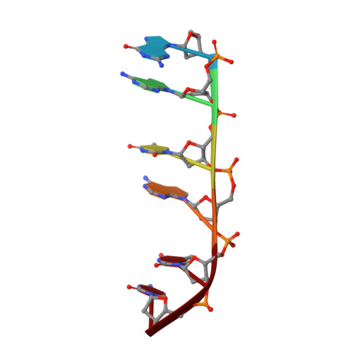
Solution Structure of a Complex between [N-Mecys3,N-Mecys7]Tandem and [D(Gatatc)]2.
Addess, K.J., Sinsheimer, J.S., Feigon, J.(1993) Biochemistry 32: 2498
- PubMed: 8448108
- DOI: https://doi.org/10.1021/bi00061a006
- Primary Citation of Related Structures:
2DA8 - PubMed Abstract:
[N-MeCys3,N-MeCys7]TANDEM (CysMeTANDEM) is an octadepsipeptide quinoxaline antibiotic that binds specifically by bisintercalation to double-stranded DNA at NTAN sites [Addess, K. J., Gilbert, D. E., Olsen, R. K., & Feigon, J. (1992) Biochemistry 31, 339-350; Addess, K. J., Gilbert, D. E., & Feigon, J. (1992) in Structure and Function Volume 1: Nucleic Acids (Sarma, R. H., & Sarma, M. H., Eds.) pp 147-164, Adenine Press, Schenectady, NY]. We have determined the three-dimensional structure of a complex of CysMeTANDEM and the DNA hexamer [d(GATATC)]2 using two-dimensional 1H NMR derived NOE and dihedral bond angle constraints. This is the first structure of a TpA-specific quinoxaline antibiotic in complex with DNA. Initial structures of the complex were generated by metric matrix distance geometry followed by simulated annealing. Eight of these structures, refined by restrained molecular dynamics, energy minimization, and NOE-based relaxation matrix refinement, have an average pairwise RMSD of 1.11 A for all structures, calculated using all heavy atoms of the drug and the DNA except the terminal base pairs. CysMeTANDEM binds to and affects the structure of the DNA in a manner similar to that observed in complexes of the CpG-specific quinoxaline antibiotics triostin A and echinomycin with DNA [Ughetto, G., Wang, A. H.-J., Quigley, G. J., van der Marel, G. A., van Boom, J. H., & Rich, A. (1985) Nucleic Acids Res. 13, 2305-2323; Wang, A. H.-J., Ughetto G., Quigley, G. J., Hakoshima, T., van der Marel, G. A., van Boom, J. H., & Rich, A. (1984) Science 225, 1115-1121; Wang, A. H.-J., Ughetto, G., Quigley, G. J., & Rich, A. (1986) J. Biomol. Struct. Dyn. 4, 319-342]. The two quinoxaline rings bisintercalate on either side of the two central T.A base pairs and the peptide ring lies in the minor groove. The central A.T base pairs of the complex are underwound (average helical twist angle of approximately -10 degrees) and buckle inward by approximately 20 degrees. There are intermolecular hydrogen bonds between each of the Ala NH and the AN3 protons of the TpA binding site, analogous to those observed between Ala NH and GN3 in the crystal structures of the CpG-specific complexes of echinomycin and triostin A with DNA. However, the structure of the peptide ring of CysMeTANDEM in the complex differs from that of echinomycin and triostin A.(ABSTRACT TRUNCATED AT 400 WORDS)
- Department of Chemistry and Biochemistry, School of Medicine, University of California, Los Angeles 90024.
Organizational Affiliation:


