2D55

Structural, physical and biological characteristics of RNA.DNA binding agent N8-actinomycin D


Experimental Data Snapshot

  • Method: X-RAY DIFFRACTION
  • Resolution: 3.00 Å
  • R-Value Work: 0.190 
  • R-Value Observed: 0.190 

wwPDB Validation   3D Report Full Report


This is version 2.0 of the entry. See complete history


Literature

Crystal Structure of the 2:1 Complex between D(Gaagcttc) and the Anticancer Drug Actinomycin D.

Kamitori, S.Takusagawa, F.

(1992) J Mol Biol 225: 445

  • DOI: https://doi.org/10.1016/0022-2836(92)90931-9
  • Primary Citation of Related Structures:  
    2D55

  • PubMed Abstract: 

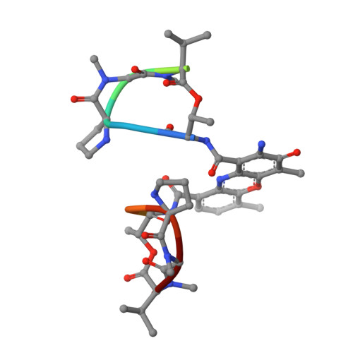
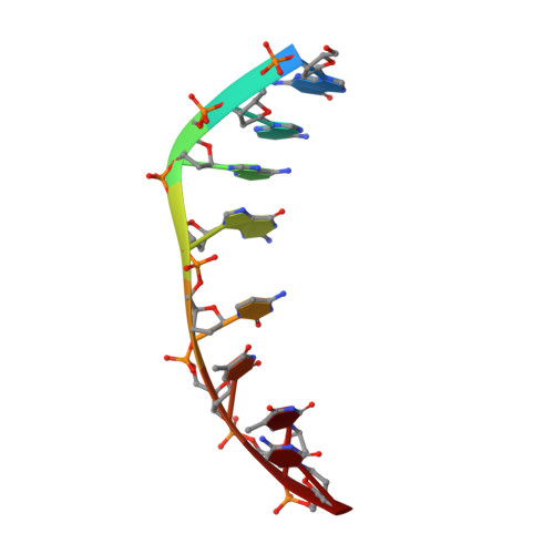
    The crystal structures of the 2:1 complex of the self-complementary DNA octamer d(GAAGCTTC) with actinomycin D has been determined at 3.0 A resolution. This is the first example of a crystal structure of a DNA-drug complex in which the drug intercalates into the middle of a relatively long DNA segment. The results finally confirmed the DNA-actinomycin intercalation model proposed by Sobell & co-workers in 1971. The DNA molecule adopts a severely distorted and slightly kinked B-DNA-like structure with an actinomycin D molecule intercalated in the middle sequence, GC. The two cyclic depsipeptides, which differ from each other in overall conformation, lie in the minor groove. The complex is further stabilized by forming base-peptide and chromophore-backbone hydrogen bonds. The DNA helix appears to be unwound by rotating one of the base-pairs at the intercalation site. This single base-pair unwinding motion generates a unique asymmetrically wound helix at the binding site of the drug, i.e. the helix is loosened at one end of the intercalation site and tightened at the other end. The large unwinding of the DNA by the drug intercalation is absorbed mostly in a few residues adjacent to the intercalation site. The asymmetrical twist of the DNA helix, the overall conformation of the two cyclic depsipeptides and their interaction mode with DNA are correlated to each other and rationally explained.


  • Organizational Affiliation

    X-ray Crystallography Laboratory, University of Kansas, Lawrence 66045-0046.


Macromolecules

Find similar proteins by:  Sequence   |   3D Structure  

Entity ID: 2
MoleculeChains Sequence LengthOrganismDetailsImage
ACTINOMYCIN D11Streptomyces antibioticusMutation(s): 0 
Entity Groups  
Sequence Clusters30% Identity50% Identity70% Identity90% Identity95% Identity100% Identity
Sequence Annotations
Expand
  • Reference Sequence

Find similar nucleic acids by:  Sequence   |   3D Structure  

Entity ID: 1
MoleculeChains LengthOrganismImage
DNA (5'-D(*GP*AP*AP*GP*CP*TP*TP*C)-3')
A, B
8N/A
Sequence Annotations
Expand
  • Reference Sequence
Small Molecules
Modified Residues  2 Unique
IDChains TypeFormula2D DiagramParent
MVA
Query on MVA
C
L-PEPTIDE LINKINGC6 H13 N O2VAL
SAR
Query on SAR
C
PEPTIDE LINKINGC3 H7 N O2GLY
Binding Affinity Annotations 
IDSourceBinding Affinity
PXZ PDBBind:  2D55 IC50: 35 (nM) from 1 assay(s)
Biologically Interesting Molecules (External Reference) 1 Unique
Experimental Data & Validation

Experimental Data

  • Method: X-RAY DIFFRACTION
  • Resolution: 3.00 Å
  • R-Value Work: 0.190 
  • R-Value Observed: 0.190 
  • Space Group: C 1 2 1
Unit Cell:
Length ( Å )Angle ( ˚ )
a = 51.37α = 90
b = 62.09β = 113.8
c = 25.04γ = 90
Software Package:
Software NamePurpose
X-PLORrefinement

Structure Validation

View Full Validation Report


Entry History 

Deposition Data

Revision History  (Full details and data files)

  • Version 1.0: 1995-10-15
    Type: Initial release
  • Version 1.1: 2011-06-14
    Changes: Version format compliance
  • Version 1.2: 2011-07-13
    Changes: Version format compliance
  • Version 1.3: 2011-07-27
    Changes: Atomic model, Database references, Derived calculations, Non-polymer description, Structure summary
  • Version 1.4: 2012-12-12
    Changes: Other
  • Version 2.0: 2023-11-15
    Changes: Advisory, Atomic model, Data collection, Database references, Derived calculations