Formaldehyde cross-links daunorubicin and DNA efficiently: HPLC and X-ray diffraction studies.
Wang, A.H., Gao, Y.G., Liaw, Y.C., Li, Y.K.(1991) Biochemistry 30: 3812-3815
- PubMed: 2018756 Search on PubMed
- DOI: https://doi.org/10.1021/bi00230a002
- Primary Citation Related Structures:
1D33, 2D34 - PubMed Abstract:
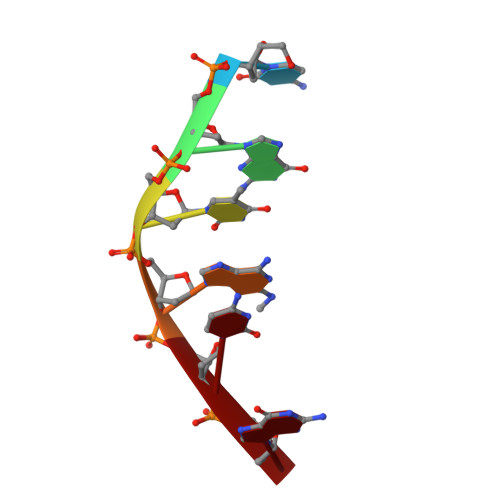
Formaldehyde (HCHO) cross-links the anticancer drug daunorubicin (DAU) to DNA efficiently. When DAU is mixed with DNA hexamers, d(CGCGCG) and d(CGTDCG), in the presence of HCHO, stable covalent adducts of DNA are formed, as shown by the HPLC analyses. The major adducts are identical with the materials in the respective crystals which can be readily obtained from the 1:1 mixture of DAU-d(CGCGCG) and DAU-d(CGTDCG) plus HCHO, but not from the solution without HCHO. The high-resolution (1.5 A) X-ray crystal structure of those adducts shows unambiguously that they contain a covalent methylene bridge between the N3' of daunosamine and the N2 of the guanine or 2-aminoadenine. The perfect juxtaposition of the two amino groups in the minor groove of the complex provides a template for an efficient addition of HCHO. The methylene bridge does not perturb the conformation of the drug-DNA complex, when compared to the structure of DAU-d(CGTACG). The results suggest new approaches for synthesizing a new type of potential anticancer drug by attaching a reactive (e.g., alkylating) functional group at the N3' amino position of daunorubicin/doxorubicin. The stable drug-DNA adduct may be useful as probes for other biological studies.
- Department of Physiology and Biophysics, University of Illinois, Urbana-Champaign 61801.
Organizational Affiliation: