2D2Z | pdb_00002d2z

Crystal structure of Soluble Form Of CLIC4


Experimental Data Snapshot

  • Method: X-RAY DIFFRACTION
  • Resolution: 2.20 Å
  • R-Value Free: 
    0.262 (Depositor), 0.254 (DCC) 
  • R-Value Work: 
    0.216 (Depositor), 0.212 (DCC) 
  • R-Value Observed: 
    0.216 (Depositor) 

wwPDB Validation 3D Report Full Report

Validation slider image for 2D2Z

This is version 1.4 of the entry. See complete history

Literature

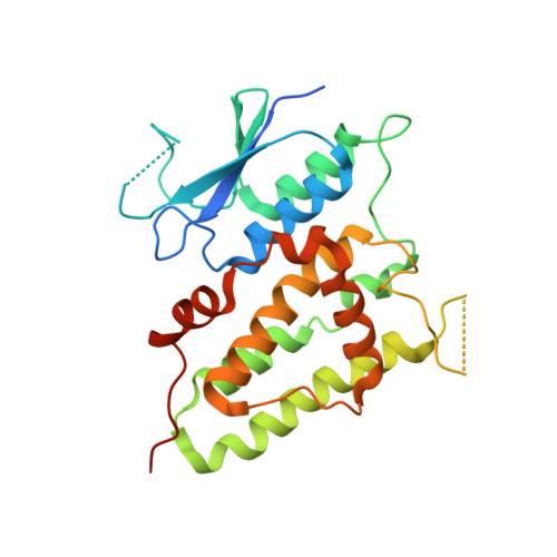
Trimeric structure of the wild soluble chloride intracellular ion channel CLIC4 observed in crystals

Li, Y.F.Li, D.F.Zeng, Z.H.Wang, D.C.

(2006) Biochem Biophys Res Commun 343: 1272-1278

  • DOI: https://doi.org/10.1016/j.bbrc.2006.03.099
  • Primary Citation Related Structures: 
    2D2Z

  • PubMed Abstract: 

    The crystal structure of a wild type of the human soluble chloride intracellular ion channel CLIC4 (wCLIC4) has been determined at a resolution of 2.2A. The structure shows a homotrimer in an asymmetric unit, which is first observed in CLICs. The assembly of the trimer takes a unique triple interaction mode between three monomers with a hydrogen-bond network and hydrophobic contacts. Through such complicated interactions, the homotrimer of wCLIC4 is firmly stabilized. The structure shows an oligomeric mode with a unique assembly mechanism by which the oligomerization of CLIC4 can be performed without any intramolecular disulfide bond formation. It indicated a possibility that CLIC4 may take a unique structural organization distinct from CLIC1 for docking with lipid bilayers. In addition, the structure shows distinct conformational states of the h2 region for respective monomers of the trimer, which reveal an intrinsic conformational susceptibility for this significant region in the structural transition.


  • Organizational Affiliation
    • National Laboratory of Biomacromolecules, Institute of Biophysics, Chinese Academy of Sciences, Beijing 100101, People's Republic of China.

Macromolecule Content 

  • Total Structure Weight: 89.64 kDa 
  • Atom Count: 6,238 
  • Modeled Residue Count: 694 
  • Deposited Residue Count: 783 
  • Unique protein chains: 1

Macromolecules

Find similar proteins by:|  3D Structure
Entity ID: 1
MoleculeChains  Sequence LengthOrganismDetailsImage
Chloride intracellular channel protein 4
A, B, C
261Homo sapiensMutation(s): 0 
EC: 1.8
UniProt & NIH Common Fund Data Resources
Find proteins for Q9Y696 (Homo sapiens)
Explore Q9Y696 
Go to UniProtKB:  Q9Y696
PHAROS:  Q9Y696
GTEx:  ENSG00000169504 
Entity Groups
Sequence Clusters30% Identity50% Identity70% Identity90% Identity95% Identity100% Identity
UniProt GroupQ9Y696
Sequence Annotations
Expand
Reference Sequence

Experimental Data & Validation

Experimental Data

  • Method: X-RAY DIFFRACTION
  • Resolution: 2.20 Å
  • R-Value Free:  0.262 (Depositor), 0.254 (DCC) 
  • R-Value Work:  0.216 (Depositor), 0.212 (DCC) 
  • R-Value Observed: 0.216 (Depositor) 
Space Group: P 1 21 1
Unit Cell:
Length ( Å )Angle ( ˚ )
a = 73.19α = 90
b = 86.05β = 112.99
c = 73.38γ = 90
Software Package:
Software NamePurpose
crystaldata collection
SCALAdata scaling
CCP4model building
CNSrefinement
CrystalCleardata reduction
CCP4data scaling
CCP4phasing

Structure Validation

View Full Validation Report


Entry History 

Deposition Data

Revision History  (Full details and data files)

  • Version 1.0: 2006-05-16
    Type: Initial release
  • Version 1.1: 2008-04-30
    Changes: Version format compliance
  • Version 1.2: 2011-07-13
    Changes: Version format compliance
  • Version 1.3: 2017-10-11
    Changes: Refinement description
  • Version 1.4: 2024-03-13
    Changes: Data collection, Database references