Improving the catalytic efficiency of a meta-cleavage product hydrolase (CumD) from Pseudomonas fluorescens IP01
Jun, S.Y., Fushinobu, S., Nojiri, H., Omori, T., Shoun, H., Wakagi, T.(2006) Biochim Biophys Acta 1764: 1159-1166
- PubMed: 16844437 Search on PubMed
- DOI: https://doi.org/10.1016/j.bbapap.2006.05.010
- Primary Citation Related Structures:
2D0D - PubMed Abstract:
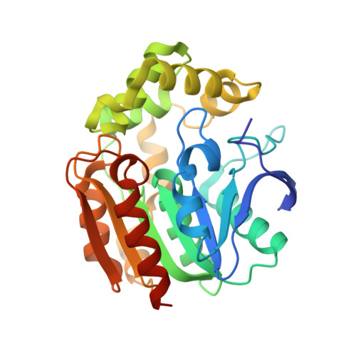
The meta-cleavage product hydrolase from Pseudomonas fluorescens IP01 (CumD) hydrolyzes 2-hydroxy-6-oxo-7-methylocta-2,4-dienoate (6-isopropyl HODA) in the cumene (isopropylbenzene) degradation pathway. To modulate the substrate specificity and catalytic efficiency of CumD toward substrates derived from monocyclic aromatic compounds, we constructed the CumD mutants, A129V, I199V, and V227I, as well as four types of double and triple mutants. Toward substrates with smaller side chains (e.g. 2-hydroxy-6-oxohepta-2,4-dienoate; 6-ethyl-HODA), the k(cat)/K(m) values of the single mutants were 4.2-11 fold higher than that of the wild type enzyme and 1.8-4.7 fold higher than that of the meta-cleavage product hydrolase from Pseudomonas putida F1 (TodF). The A129V mutant showed the highest k(cat)/K(m) value for 2-hydroxy-6-oxohepta-2,4-dienoate (6-ethyl-HODA). The crystal structure of the A129V mutant was determined at 1.65 A resolution, enabling location of the Ogamma atom of the Ser103 side chain. A chloride ion was bound to the oxyanion hole of the active site, and mutant enzymes at the residues forming this site were also examined. The k(cat) values of Ser34 mutants were decreased 2.9-65 fold, suggesting that the side chain of Ser34 supports catalysis by stabilizing the anionic oxygen of the proposed intermediate state (gem-diolate). This is the first crystal structure determination of CumD in an active form, with the Ser103 residue, one of the catalytically essential "triad", being intact.
- Laboratory of Enzymology, Department of Biotechnology, The University of Tokyo, 1-1-1 Yayoi, Bunkyo-ku, Tokyo 113-8657, Japan.
Organizational Affiliation: