2CY7 | pdb_00002cy7

The crystal structure of human Atg4B


Experimental Data Snapshot

  • Method: X-RAY DIFFRACTION
  • Resolution: 1.90 Å
  • R-Value Free: 
    0.247 (Depositor), 0.246 (DCC) 
  • R-Value Work: 
    0.215 (Depositor), 0.215 (DCC) 
  • R-Value Observed: 
    0.215 (Depositor) 

wwPDB Validation 3D Report Full Report

Validation slider image for 2CY7

This is version 1.3 of the entry. See complete history

Literature

Structural Basis for the Specificity and Catalysis of Human Atg4B Responsible for Mammalian Autophagy

Sugawara, K.Suzuki, N.N.Fujioka, Y.Mizushima, N.Ohsumi, Y.Inagaki, F.

(2005) J Biological Chem 280: 40058-40065

  • DOI: https://doi.org/10.1074/jbc.M509158200
  • Primary Citation Related Structures: 
    2CY7

  • PubMed Abstract: 

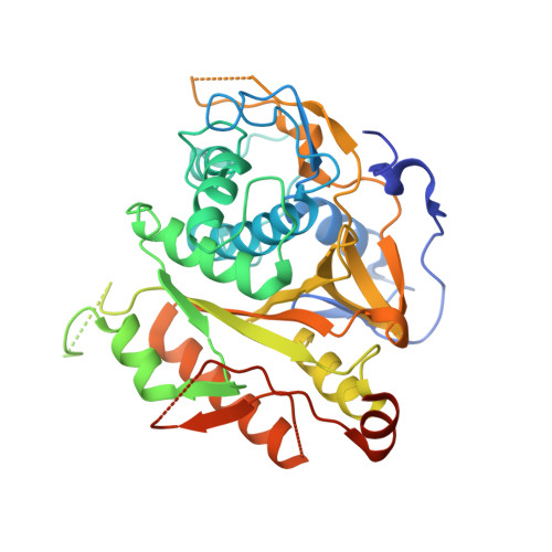
    Reversible modification of Atg8 with phosphatidylethanolamine is crucial for autophagy, the bulk degradation system conserved in eukaryotic cells. Atg4 is a novel cysteine protease that processes and deconjugates Atg8. Herein, we report the crystal structure of human Atg4B (HsAtg4B) at 1.9-A resolution. Despite no obvious sequence homology with known proteases, the structure of HsAtg4B shows a classical papain-like fold. In addition to the papain fold region, HsAtg4B has a small alpha/beta-fold domain. This domain is thought to be the binding site for Atg8 homologs. The active site cleft of HsAtg4B is masked by a loop (residues 259-262), implying a conformational change upon substrate binding. The structure and in vitro mutational analyses provide the basis for the specificity and catalysis of HsAtg4B. This will enable the design of Atg4-specific inhibitors that block autophagy.


  • Organizational Affiliation
    • Department of Structural Biology, Graduate School of Pharmaceutical Sciences, Hokkaido University, N-12, W-6, Kita-ku, Sapporo 060-0812, Japan.

Macromolecule Content 

  • Total Structure Weight: 44.65 kDa 
  • Atom Count: 2,817 
  • Modeled Residue Count: 333 
  • Deposited Residue Count: 396 
  • Unique protein chains: 1

Macromolecules

Find similar proteins by:|  3D Structure
Entity ID: 1
MoleculeChains  Sequence LengthOrganismDetailsImage
Cysteine protease APG4B396Homo sapiensMutation(s): 0 
EC: 3.4.22
UniProt & NIH Common Fund Data Resources
Find proteins for Q9Y4P1 (Homo sapiens)
Explore Q9Y4P1 
Go to UniProtKB:  Q9Y4P1
PHAROS:  Q9Y4P1
GTEx:  ENSG00000168397 
Entity Groups
Sequence Clusters30% Identity50% Identity70% Identity90% Identity95% Identity100% Identity
UniProt GroupQ9Y4P1
Sequence Annotations
Expand
Reference Sequence

Experimental Data & Validation

Experimental Data

  • Method: X-RAY DIFFRACTION
  • Resolution: 1.90 Å
  • R-Value Free:  0.247 (Depositor), 0.246 (DCC) 
  • R-Value Work:  0.215 (Depositor), 0.215 (DCC) 
  • R-Value Observed: 0.215 (Depositor) 
Space Group: C 2 2 21
Unit Cell:
Length ( Å )Angle ( ˚ )
a = 51.913α = 90
b = 88.339β = 90
c = 160.139γ = 90
Software Package:
Software NamePurpose
CNSrefinement
HKL-2000data reduction
SCALEPACKdata scaling
SOLVEphasing

Structure Validation

View Full Validation Report


Entry History 

Deposition Data

Revision History  (Full details and data files)

  • Version 1.0: 2005-09-13
    Type: Initial release
  • Version 1.1: 2008-04-30
    Changes: Version format compliance
  • Version 1.2: 2011-07-13
    Changes: Version format compliance
  • Version 1.3: 2024-03-13
    Changes: Data collection, Database references