Structural and Functional Differences of SWIRM Domain Subtypes
Yoneyama, M., Tochio, N., Umehara, T., Koshiba, S., Inoue, M., Yabuki, T., Aoki, M., Seki, E., Matsuda, T., Watanabe, S., Tomo, Y., Nishimura, Y., Harada, T., Terada, T., Shirouzu, M., Hayashizaki, Y., Ohara, O., Tanaka, A., Kigawa, T., Yokoyama, S.(2007) J Mol Biology 369: 222-238
- PubMed: 17428495 Search on PubMed
- DOI: https://doi.org/10.1016/j.jmb.2007.03.027
- Primary Citation Related Structures:
2CU7, 2CUJ, 2DCE - PubMed Abstract:
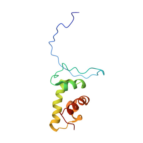
SWIRM is a conserved domain found in several chromatin-associated proteins. Based on their sequences, the SWIRM family members can be classified into three subfamilies, which are represented by Swi3, LSD1, and Ada2. Here we report the SWIRM structure of human MYb-like, Swirm and Mpn domain-containing protein-1 (MYSM1). The MYSM1 SWIRM structure forms a compact HTH-related fold comprising five alpha-helices, which best resembles the Swi3 SWIRM structure, among the known SWIRM structures. The MYSM1 and Swi3 SWIRM structures are more similar to the LSD1 structure than the Ada2alpha structure. The SWIRM domains of MYSM1 and LSD1 lacked DNA binding activity, while those of Ada2alpha and the human Swi3 counterpart, SMARCC2, bound DNA. The dissimilarity in the DNA-binding ability of the MYSM1 and SMARCC2 SWIRM domains might be due to a couple of amino acid differences in the last helix. These results indicate that the SWIRM family has indeed diverged into three structural subfamilies (Swi3/MYSM1, LSD1, and Ada2 types), and that the Swi3/MYSM1-type subfamily has further diverged into two functionally distinct groups. We also solved the structure of the SANT domain of MYSM1, and demonstrated that it bound DNA with a similar mode to that of the c-Myb DNA-binding domain.
- RIKEN Genomic Sciences Center, 1-7-22 Suehiro-cho, Tsurumi, Yokohama 230-0045, Japan.
Organizational Affiliation: