2CRK | pdb_00002crk

MUSCLE CREATINE KINASE


Experimental Data Snapshot

  • Method: X-RAY DIFFRACTION
  • Resolution: 2.35 Å
  • R-Value Free: 
    0.288 (Depositor) 
  • R-Value Work: 
    0.195 (Depositor) 
  • R-Value Observed: 
    0.195 (Depositor) 

Starting Model: experimental
View more details

wwPDB Validation 3D Report Full Report

Validation slider image for 2CRK

This is version 1.3 of the entry. See complete history

Literature

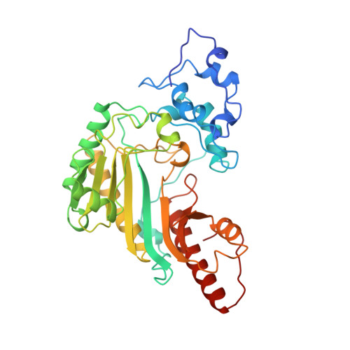
Crystal structure of rabbit muscle creatine kinase.

Rao, J.K.Bujacz, G.Wlodawer, A.

(1998) FEBS Lett 439: 133-137

  • DOI: https://doi.org/10.1016/s0014-5793(98)01355-6
  • Primary Citation Related Structures: 
    2CRK

  • PubMed Abstract: 

    The crystal structure of rabbit muscle creatine kinase, solved at 2.35 A resolution by X-ray diffraction methods, clearly identified the active site with bound sulfates surrounded by a constellation of arginine residues. The putative binding site of creatine, which is occupied by a sulfate group in this analysis, has been tentatively identified. The dimeric interface of the enzyme is held together by a small number of hydrogen bonds.


  • Organizational Affiliation
    • Macromolecular Structure Laboratory, NCI-Frederick Cancer Research and Development Center, ABL-Basic Research Program, Frederick, MD 21702, USA. rao@ncifcrf.gov

Macromolecule Content 

  • Total Structure Weight: 43.69 kDa 
  • Atom Count: 3,379 
  • Modeled Residue Count: 365 
  • Deposited Residue Count: 381 
  • Unique protein chains: 1

Macromolecules

Find similar proteins by:|  3D Structure
Entity ID: 1
MoleculeChains  Sequence LengthOrganismDetailsImage
PROTEIN (CREATINE KINASE)381Oryctolagus cuniculusMutation(s): 0 
EC: 2.7.3.2
UniProt
Find proteins for P00563 (Oryctolagus cuniculus)
Explore P00563 
Go to UniProtKB:  P00563
Entity Groups
Sequence Clusters30% Identity50% Identity70% Identity90% Identity95% Identity100% Identity
UniProt GroupP00563
Sequence Annotations
Expand
Reference Sequence

Experimental Data & Validation

Experimental Data

  • Method: X-RAY DIFFRACTION
  • Resolution: 2.35 Å
  • R-Value Free:  0.288 (Depositor) 
  • R-Value Work:  0.195 (Depositor) 
  • R-Value Observed: 0.195 (Depositor) 
Space Group: I 4 2 2
Unit Cell:
Length ( Å )Angle ( ˚ )
a = 199.6α = 90
b = 199.6β = 90
c = 71γ = 90
Software Package:
Software NamePurpose
PHASESphasing
AMoREphasing
MADIRAmodel building
X-PLORrefinement
DENZOdata reduction
SCALEPACKdata scaling
MADIRAphasing

Structure Validation

View Full Validation Report


Entry History 

Deposition Data

Revision History  (Full details and data files)

  • Version 1.0: 1999-01-13
    Type: Initial release
  • Version 1.1: 2007-10-16
    Changes: Version format compliance
  • Version 1.2: 2011-07-13
    Changes: Version format compliance
  • Version 1.3: 2023-08-23
    Changes: Data collection, Database references, Derived calculations, Refinement description