2CNB | pdb_00002cnb

Trypanosoma brucei UDP-galactose-4-epimerase in ternary complex with NAD and the substrate analogue UDP-4-deoxy-4-fluoro-alpha-D-galactose


Experimental Data Snapshot

  • Method: X-RAY DIFFRACTION
  • Resolution: 2.70 Å
  • R-Value Free: 
    0.281 (Depositor), 0.280 (DCC) 
  • R-Value Work: 
    0.199 (Depositor), 0.200 (DCC) 
  • R-Value Observed: 
    0.203 (Depositor) 

wwPDB Validation   3D Report Full Report


Ligand Structure Quality Assessment 

Created with Raphaël 2.3.0Worse 01 BetterLigand structure goodness of fit to experimental dataBest fitted NADClick on this verticalbar to view detailsBest fitted UFGClick on this verticalbar to view details

This is version 1.3 of the entry. See complete history


Literature

Trypanosoma Brucei Udp-Galactose-4-Epimerase in Ternary Complex with Nad+ and the Substrate Analogue Udp-4-Deoxy-4-Fluoro-Alpha-D-Galactose

Alphey, M.S.Burton, A.Urbaniak, M.Boons, G.J.Ferguson, M.A.J.Hunter, W.N.

(2006) Acta Crystallogr Sect F Struct Biol Cryst Commun 62: 829

  • DOI: https://doi.org/10.1107/S1744309106028740
  • Primary Citation of Related Structures:  
    2CNB

  • PubMed Abstract: 

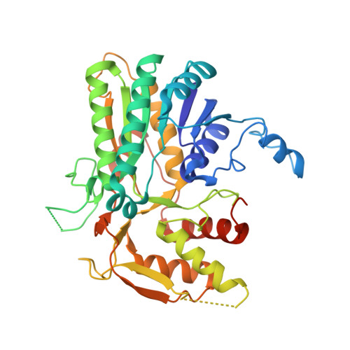
    The structure of the NAD-dependent oxidoreductase UDP-galactose-4'-epimerase from Trypanosoma brucei in complex with cofactor and the substrate analogue UDP-4-deoxy-4-fluoro-alpha-D-galactose has been determined using diffraction data to 2.7 A resolution. Despite the high level of sequence and structure conservation between the trypanosomatid enzyme and those from humans, yeast and bacteria, the binding of the 4-fluoro-alpha-D-galactose moiety is distinct from previously reported structures. Of particular note is the observation that when bound to the T. brucei enzyme, the galactose moiety of this fluoro-derivative is rotated approximately 180 degrees with respect to the orientation of the hexose component of UDP-glucose when in complex with the human enzyme. The architecture of the catalytic centre is designed to effectively bind different orientations of the hexose, a finding that is consistent with a mechanism that requires the sugar to maintain a degree of flexibility within the active site.


  • Organizational Affiliation

    Division of Biological Chemistry and Molecular Microbiology, School of Life Sciences, University of Dundee, Dundee DD1 5EH, Scotland.


Macromolecules
Find similar proteins by:  (by identity cutoff)  |  3D Structure
Entity ID: 1
MoleculeChains Sequence LengthOrganismDetailsImage
UDP-GALACTOSE-4-EPIMERASE
A, B, C, D
397Trypanosoma bruceiMutation(s): 0 
EC: 5.1.3.2
UniProt
Find proteins for Q8T8E9 (Trypanosoma brucei)
Explore Q8T8E9 
Go to UniProtKB:  Q8T8E9
Entity Groups  
Sequence Clusters30% Identity50% Identity70% Identity90% Identity95% Identity100% Identity
UniProt GroupQ8T8E9
Sequence Annotations
Expand
  • Reference Sequence
Small Molecules
Ligands 2 Unique
IDChains Name / Formula / InChI Key2D Diagram3D Interactions
NAD
Query on NAD

Download Ideal Coordinates CCD File 
F [auth A],
H [auth B],
J [auth C],
L [auth D]
NICOTINAMIDE-ADENINE-DINUCLEOTIDE
C21 H27 N7 O14 P2
BAWFJGJZGIEFAR-NNYOXOHSSA-N
UFG
Query on UFG

Download Ideal Coordinates CCD File 
E [auth A],
G [auth B],
I [auth C],
K [auth D]
URIDINE-5'-DIPHOSPHATE-4-DEOXY-4-FLUORO-ALPHA-D-GALACTOSE
C15 H23 F N2 O16 P2
OAPPZHVTNHJVAL-JZMIEXBBSA-N
Experimental Data & Validation

Experimental Data

  • Method: X-RAY DIFFRACTION
  • Resolution: 2.70 Å
  • R-Value Free:  0.281 (Depositor), 0.280 (DCC) 
  • R-Value Work:  0.199 (Depositor), 0.200 (DCC) 
  • R-Value Observed: 0.203 (Depositor) 
Space Group: P 21 21 21
Unit Cell:
Length ( Å )Angle ( ˚ )
a = 101.724α = 90
b = 111.694β = 90
c = 160.873γ = 90
Software Package:
Software NamePurpose
REFMACrefinement
DENZOdata reduction
SCALEPACKdata scaling

Structure Validation

View Full Validation Report


Ligand Structure Quality Assessment 

Created with Raphaël 2.3.0Worse 01 BetterLigand structure goodness of fit to experimental dataBest fitted NADClick on this verticalbar to view detailsBest fitted UFGClick on this verticalbar to view details

Entry History 

Deposition Data

Revision History  (Full details and data files)

  • Version 1.0: 2006-06-12
    Type: Initial release
  • Version 1.1: 2011-05-08
    Changes: Version format compliance
  • Version 1.2: 2011-07-13
    Changes: Version format compliance
  • Version 1.3: 2024-05-08
    Changes: Data collection, Database references, Other