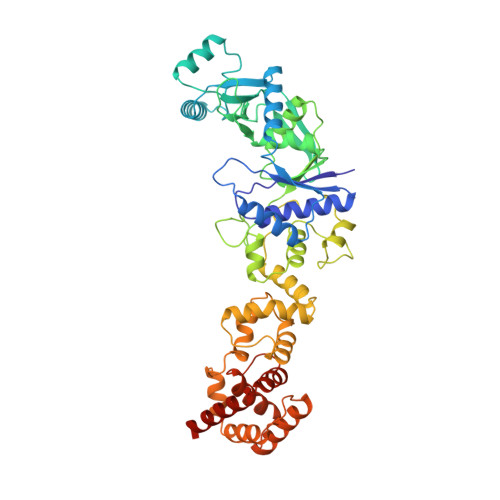
2CFO

Non-Discriminating Glutamyl-tRNA Synthetase from Thermosynechococcus elongatus in Complex with Glu


Experimental Data Snapshot

  • Method: X-RAY DIFFRACTION
  • Resolution: 2.45 Å
  • R-Value Free: 0.256 
  • R-Value Work: 0.186 
  • R-Value Observed: 0.189 

Starting Model: experimental
View more details

wwPDB Validation   3D Report Full Report


This is version 1.2 of the entry. See complete history


Literature

Crystal Structure of a Non-Discriminating Glutamyl- tRNA Synthetase.

Schulze, J.O.Masoumi, A.Nickel, D.Jahn, M.Jahn, D.Schubert, W.-D.Heinz, D.W.

(2006) J Mol Biol 361: 888

  • DOI: https://doi.org/10.1016/j.jmb.2006.06.054
  • Primary Citation of Related Structures:  
    2CFO

  • PubMed Abstract: 

    Error-free protein biosynthesis is dependent on the reliable charging of each tRNA with its cognate amino acid. Many bacteria, however, lack a glutaminyl-tRNA synthetase. In these organisms, tRNA(Gln) is initially mischarged with glutamate by a non-discriminating glutamyl-tRNA synthetase (ND-GluRS). This enzyme thus charges both tRNA(Glu) and tRNA(Gln) with glutamate. Discriminating GluRS (D-GluRS), found in some bacteria and all eukaryotes, exclusively generates Glu-tRNA(Glu). Here we present the first crystal structure of a non-discriminating GluRS from Thermosynechococcus elongatus (ND-GluRS(Tel)) in complex with glutamate at a resolution of 2.45 A. Structurally, the enzyme shares the overall architecture of the discriminating GluRS from Thermus thermophilus (D-GluRS(Tth)). We confirm experimentally that GluRS(Tel) is non-discriminating and present kinetic parameters for synthesis of Glu-tRNA(Glu) and of Glu-tRNA(Gln). Anticodons of tRNA(Glu) (34C/UUC36) and tRNA(Gln) (34C/UUG36) differ only in base 36. The pyrimidine base of C36 is specifically recognized in D-GluRS(Tth) by the residue Arg358. In ND-GluRS(Tel) this arginine residue is replaced by glycine (Gly366) presumably allowing both cytosine and the bulkier purine base G36 of tRNA(Gln) to be tolerated. Most other ND-GluRS share this structural feature, leading to relaxed substrate specificity.


  • Organizational Affiliation

    Division of Structural Biology, German Research Centre for Biotechnology (GBF), Mascheroder Weg 1, D-38124 Braunschweig, Germany.


Macromolecules
Find similar proteins by:  (by identity cutoff)  |  3D Structure
Entity ID: 1
MoleculeChains Sequence LengthOrganismDetailsImage
GLUTAMYL-TRNA SYNTHETASE
A, B
492Synechococcus elongatusMutation(s): 0 
EC: 6.1.1.17
UniProt
Find proteins for Q8DLI5 (Thermosynechococcus vestitus (strain NIES-2133 / IAM M-273 / BP-1))
Explore Q8DLI5 
Go to UniProtKB:  Q8DLI5
Entity Groups  
Sequence Clusters30% Identity50% Identity70% Identity90% Identity95% Identity100% Identity
UniProt GroupQ8DLI5
Sequence Annotations
Expand
  • Reference Sequence
Small Molecules
Ligands 1 Unique
IDChains Name / Formula / InChI Key2D Diagram3D Interactions
GLU
Query on GLU

Download Ideal Coordinates CCD File 
C [auth A]GLUTAMIC ACID
C5 H9 N O4
WHUUTDBJXJRKMK-VKHMYHEASA-N
Experimental Data & Validation

Experimental Data

  • Method: X-RAY DIFFRACTION
  • Resolution: 2.45 Å
  • R-Value Free: 0.256 
  • R-Value Work: 0.186 
  • R-Value Observed: 0.189 
  • Space Group: P 1 21 1
Unit Cell:
Length ( Å )Angle ( ˚ )
a = 36.149α = 90
b = 99.603β = 91.71
c = 182.407γ = 90
Software Package:
Software NamePurpose
REFMACrefinement
XDSdata reduction
XSCALEdata scaling
PHASERphasing

Structure Validation

View Full Validation Report


Entry History 

Deposition Data

Revision History  (Full details and data files)

  • Version 1.0: 2006-08-16
    Type: Initial release
  • Version 1.1: 2011-07-13
    Changes: Refinement description, Version format compliance
  • Version 1.2: 2023-12-13
    Changes: Data collection, Database references, Other, Refinement description