2CB1 | pdb_00002cb1

Crystal Structure of O-actetyl Homoserine Sulfhydrylase From Thermus Thermophilus HB8,OAH2.


Experimental Data Snapshot

  • Method: X-RAY DIFFRACTION
  • Resolution: 2.00 Å
  • R-Value Free: 
    0.234 (Depositor), 0.229 (DCC) 
  • R-Value Work: 
    0.187 (Depositor), 0.186 (DCC) 
  • R-Value Observed: 
    0.190 (Depositor) 

Starting Model: experimental
View more details

wwPDB Validation 3D Report Full Report

Validation slider image for 2CB1

Ligand Structure Quality Assessment 


This is version 1.2 of the entry. See complete history

Literature

The Crystal Structure of O-Acetyl Homoserine Sulfhydrylase

Imagawa, T.Utsunomiya, H.Agari, Y.Satoh, S.Tsuge, H.

To be published.

Macromolecule Content 

  • Total Structure Weight: 44.48 kDa 
  • Atom Count: 3,221 
  • Modeled Residue Count: 405 
  • Deposited Residue Count: 412 
  • Unique protein chains: 1

Macromolecules

Find similar proteins by:|  3D Structure
Entity ID: 1
MoleculeChains  Sequence LengthOrganismDetailsImage
O-ACETYL HOMOSERINE SULFHYDRYLASE412Thermus thermophilus HB8Mutation(s): 0 
EC: 4.2.99.10 (PDB Primary Data), 2.5.1 (UniProt)
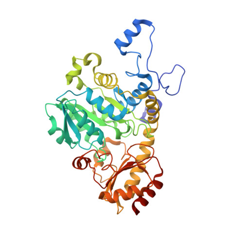
UniProt
Find proteins for Q5SJ58 (Thermus thermophilus (strain ATCC 27634 / DSM 579 / HB8))
Explore Q5SJ58 
Go to UniProtKB:  Q5SJ58
Entity Groups
Sequence Clusters30% Identity50% Identity70% Identity90% Identity95% Identity100% Identity
UniProt GroupQ5SJ58
Sequence Annotations
Expand
Reference Sequence

Small Molecules

Ligands 1 Unique
IDChains Name / Formula / InChI Key2D Diagram3D Interactions
PLP

Query on PLP


Download:Ideal Coordinates CCD File
B [auth A]PYRIDOXAL-5'-PHOSPHATE
C8 H10 N O6 P
NGVDGCNFYWLIFO-UHFFFAOYSA-N

Experimental Data & Validation

Experimental Data

  • Method: X-RAY DIFFRACTION
  • Resolution: 2.00 Å
  • R-Value Free:  0.234 (Depositor), 0.229 (DCC) 
  • R-Value Work:  0.187 (Depositor), 0.186 (DCC) 
  • R-Value Observed: 0.190 (Depositor) 
Space Group: I 2 2 2
Unit Cell:
Length ( Å )Angle ( ˚ )
a = 55.986α = 90
b = 86.392β = 90
c = 156.138γ = 90
Software Package:
Software NamePurpose
REFMACrefinement
HKL-2000data reduction
SCALEPACKdata scaling
MOLREPphasing

Structure Validation

View Full Validation Report


Ligand Structure Quality Assessment 


Entry History 

Deposition Data

Revision History  (Full details and data files)

  • Version 1.0: 2007-01-03
    Type: Initial release
  • Version 1.1: 2015-12-16
    Changes: Atomic model, Database references, Derived calculations, Non-polymer description, Other, Refinement description, Source and taxonomy, Structure summary, Version format compliance
  • Version 1.2: 2023-12-13
    Changes: Data collection, Database references, Derived calculations, Other, Refinement description