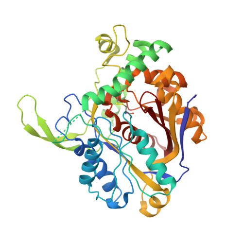
The Crystal Structure of a Plant 3-Ketoacyl-Coa Thiolase Reveals the Potential for Redox Control of Peroxisomal Fatty Acid Beta-Oxidation.
Sundaramoorthy, R., Micossi, E., Alphey, M.S., Germain, V., Bryce, J.H., Smith, S.M., Leonard, G.A., Hunter, W.N.(2006) J Mol Biology 359: 347
- PubMed: 16630629 Search on PubMed
- DOI: https://doi.org/10.1016/j.jmb.2006.03.032
- Primary Citation Related Structures:
2C7Y, 2C7Z - PubMed Abstract:
Crystal structures of peroxisomal Arabidopsis thaliana 3-ketoacyl-CoA thiolase (AtKAT), an enzyme of fatty acid beta-oxidation, are reported. The subunit, a typical thiolase, is a combination of two similar alpha/beta domains capped with a loop domain. The comparison of AtKAT with the Saccharomyces cerevisiae homologue (ScKAT) structure reveals a different placement of subunits within the functional dimers and that a polypeptide segment forming an extended loop around the open catalytic pocket of ScKAT converts to alpha-helix in AtKAT, and occludes the active site. A disulfide is formed between Cys192, on this helix, and Cys138, a catalytic residue. Access to Cys138 is determined by the structure of this polypeptide segment. AtKAT represents an oxidized, previously unknown inactive form, whilst ScKAT is the reduced and active enzyme. A high level of sequence conservation is observed, including Cys192, in eukaryotic peroxisomal, but not mitochondrial or prokaryotic KAT sequences, for this labile loop/helix segment. This indicates that KAT activity in peroxisomes is influenced by a disulfide/dithiol change linking fatty acid beta-oxidation with redox regulation.
- Division of Biological Chemistry and Molecular Microbiology, School of Life Sciences, University of Dundee, Dundee DD1 5EH, UK.
Organizational Affiliation: