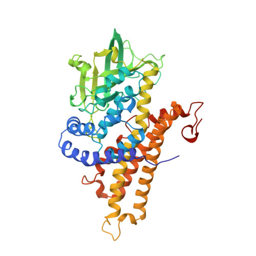
2C12

Crystal Structure of Nitroalkane Oxidase in Complex with Spermine, a Competitive Inhibitor


Experimental Data Snapshot

  • Method: X-RAY DIFFRACTION
  • Resolution: 2.07 Å
  • R-Value Free: 0.225 
  • R-Value Work: 0.188 
  • R-Value Observed: 0.190 

Starting Model: experimental
View more details

wwPDB Validation   3D Report Full Report


Ligand Structure Quality Assessment 


This is version 1.2 of the entry. See complete history


Literature

Crystal Structures of Nitroalkane Oxidase: Insights Into the Reaction Mechanism from a Covalent Complex of the Flavoenzyme Trapped During Turnover.

Nagpal, A.Valley, M.P.Fitzpatrick, P.F.Orville, A.M.

(2006) Biochemistry 45: 1138

  • DOI: https://doi.org/10.1021/bi051966w
  • Primary Citation of Related Structures:  
    2C0U, 2C12

  • PubMed Abstract: 

    Nitroalkane oxidase (NAO) from Fusarium oxysporum catalyzes the oxidation of neutral nitroalkanes to the corresponding aldehydes or ketones with the production of H(2)O(2) and nitrite. The flavoenzyme is a new member of the acyl-CoA dehydrogenase (ACAD) family, but it does not react with acyl-CoA substrates. We present the 2.2 A resolution crystal structure of NAO trapped during the turnover of nitroethane as a covalent N5-FAD adduct (ES*). The homotetrameric structure of ES* was solved by MAD phasing with 52 Se-Met sites in an orthorhombic space group. The electron density for the N5-(2-nitrobutyl)-1,5-dihydro-FAD covalent intermediate is clearly resolved. The structure of ES was used to solve the crystal structure of oxidized NAO at 2.07 A resolution. The c axis for the trigonal space group of oxidized NAO is 485 A, and there are six subunits (1(1)/(2) holoenzymes) in the asymmetric unit. Four of the active sites contain spermine (EI), a weak competitive inhibitor, and two do not contain spermine (E(ox)). The active-site structures of E(ox), EI, and ES* reveal a hydrophobic channel that extends from the exterior of the protein and terminates at Asp402 and the N5 position on the re face of the FAD. Thus, Asp402 is in the correct position to serve as the active-site base, where it is proposed to abstract the alpha proton from neutral nitroalkane substrates. The structures for NAO and various members of the ACAD family overlay with root-mean-square deviations between 1.7 and 3.1 A. The homologous region typically spans more than 325 residues and includes Glu376, which is the active-site base in the prototypical member of the ACAD family. However, NAO and the ACADs exhibit differences in hydrogen-bonding patterns between the respective active-site base, substrate molecules, and FAD. These likely differentiate NAO from the homologues and, consequently, are proposed to result in the unique reaction mechanism of NAO.


  • Organizational Affiliation

    School of Chemistry and Biochemistry, Georgia Institute of Technology, Atlanta, Georgia 30332-0400, USA.


Macromolecules
Find similar proteins by:  (by identity cutoff)  |  3D Structure
Entity ID: 1
MoleculeChains Sequence LengthOrganismDetailsImage
NITROALKANE OXIDASE
A, B, C, D, E
A, B, C, D, E, F
439Fusarium oxysporumMutation(s): 0 
EC: 1.7.3.1
UniProt
Find proteins for Q8X1D8 (Fusarium oxysporum)
Explore Q8X1D8 
Go to UniProtKB:  Q8X1D8
Entity Groups  
Sequence Clusters30% Identity50% Identity70% Identity90% Identity95% Identity100% Identity
UniProt GroupQ8X1D8
Sequence Annotations
Expand
  • Reference Sequence
Small Molecules
Ligands 4 Unique
IDChains Name / Formula / InChI Key2D Diagram3D Interactions
FAD
Query on FAD

Download Ideal Coordinates CCD File 
DA [auth F]
G [auth A]
I [auth B]
O [auth C]
U [auth D]
DA [auth F],
G [auth A],
I [auth B],
O [auth C],
U [auth D],
Z [auth E]
FLAVIN-ADENINE DINUCLEOTIDE
C27 H33 N9 O15 P2
VWWQXMAJTJZDQX-UYBVJOGSSA-N
PE4
Query on PE4

Download Ideal Coordinates CCD File 
GA [auth F],
M [auth B]
2-{2-[2-(2-{2-[2-(2-ETHOXY-ETHOXY)-ETHOXY]-ETHOXY}-ETHOXY)-ETHOXY]-ETHOXY}-ETHANOL
C16 H34 O8
PJWQOENWHPEPKI-UHFFFAOYSA-N
SPM
Query on SPM

Download Ideal Coordinates CCD File 
EA [auth F],
J [auth B],
P [auth C],
V [auth D]
SPERMINE
C10 H26 N4
PFNFFQXMRSDOHW-UHFFFAOYSA-N
GOL
Query on GOL

Download Ideal Coordinates CCD File 
AA [auth E]
BA [auth E]
CA [auth E]
FA [auth F]
H [auth A]
AA [auth E],
BA [auth E],
CA [auth E],
FA [auth F],
H [auth A],
K [auth B],
L [auth B],
N [auth C],
Q [auth C],
R [auth C],
S [auth C],
T [auth D],
W [auth D],
X [auth D],
Y [auth D]
GLYCEROL
C3 H8 O3
PEDCQBHIVMGVHV-UHFFFAOYSA-N
Experimental Data & Validation

Experimental Data

  • Method: X-RAY DIFFRACTION
  • Resolution: 2.07 Å
  • R-Value Free: 0.225 
  • R-Value Work: 0.188 
  • R-Value Observed: 0.190 
  • Space Group: P 32 2 1
Unit Cell:
Length ( Å )Angle ( ˚ )
a = 103.393α = 90
b = 103.393β = 90
c = 485.134γ = 120
Software Package:
Software NamePurpose
REFMACrefinement
HKL-2000data reduction
SCALEPACKdata scaling
MOLREPphasing

Structure Validation

View Full Validation Report


Ligand Structure Quality Assessment 


Entry History 

Deposition Data

Revision History  (Full details and data files)

  • Version 1.0: 2006-02-01
    Type: Initial release
  • Version 1.1: 2013-12-04
    Changes: Derived calculations, Non-polymer description, Other, Source and taxonomy, Version format compliance
  • Version 1.2: 2023-12-13
    Changes: Data collection, Database references, Derived calculations, Other, Refinement description