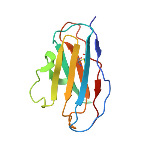
2BX5

Is FR1 the antibody's Achillies heel


Experimental Data Snapshot

  • Method: X-RAY DIFFRACTION
  • Resolution: 2.70 Å
  • R-Value Free: 0.290 
  • R-Value Work: 0.250 
  • R-Value Observed: 0.250 

Starting Model: experimental
View more details

wwPDB Validation   3D Report Full Report


This is version 1.3 of the entry. See complete history


Literature

Beta-Edge Interactions in a Pentadecameric Human Antibody Vkappa Domain.

James, L.C.Jones, P.C.Mccoy, A.Tennent, G.A.Pepys, M.B.Famm, K.Winter, G.

(2007) J Mol Biol 367: 603

  • DOI: https://doi.org/10.1016/j.jmb.2006.10.093
  • Primary Citation of Related Structures:  
    2BX5

  • PubMed Abstract: 

    Antibodies are the archetypal molecules of the Ig-fold superfamily. Their highly conserved beta-sheet architecture has evolved to avoid aggregation by protecting edge strands. However, the crystal structure of a human V kappa domain described here, reveals an exposed beta-edge strand which mediates assembly of a helical pentadecameric oligomer. This edge strand is highly conserved in V kappa domains but is both shortened and capped by the use of two sequential trans-proline residues in V lambda domains. We suggest that the exposure of this beta-edge in V kappa domains may explain why light-chain deposition disease is mediated predominantly by kappa antibodies.


  • Organizational Affiliation

    MRC Laboratory of Molecular Biology, Hills Road, Cambridge, CB2 2QH, UK. lcj@mrc-lmb.cam.ac.uk


Macromolecules
Find similar proteins by:  (by identity cutoff)  |  3D Structure
Entity ID: 1
MoleculeChains Sequence LengthOrganismDetailsImage
VD9 VKI LIGHT-CHAIN
A, B, C, D, E
A, B, C, D, E, F, G, H, I, J, K, L, M, N, O
107Homo sapiensMutation(s): 0 
UniProt
Find proteins for Q9UL77 (Homo sapiens)
Explore Q9UL77 
Go to UniProtKB:  Q9UL77
Entity Groups  
Sequence Clusters30% Identity50% Identity70% Identity90% Identity95% Identity100% Identity
UniProt GroupQ9UL77
Sequence Annotations
Expand
  • Reference Sequence
Experimental Data & Validation

Experimental Data

  • Method: X-RAY DIFFRACTION
  • Resolution: 2.70 Å
  • R-Value Free: 0.290 
  • R-Value Work: 0.250 
  • R-Value Observed: 0.250 
  • Space Group: P 64 2 2
Unit Cell:
Length ( Å )Angle ( ˚ )
a = 191.928α = 90
b = 191.928β = 90
c = 197.439γ = 120
Software Package:
Software NamePurpose
REFMACrefinement
PHASERphasing

Structure Validation

View Full Validation Report


Entry History 

Deposition Data

  • Released Date: 2006-11-15 
  • Deposition Author(s): James, L.C.

Revision History  (Full details and data files)

  • Version 1.0: 2006-11-15
    Type: Initial release
  • Version 1.1: 2013-10-30
    Changes: Derived calculations, Other, Structure summary, Version format compliance
  • Version 1.2: 2014-01-08
    Changes: Source and taxonomy
  • Version 1.3: 2023-12-13
    Changes: Data collection, Database references, Other, Refinement description