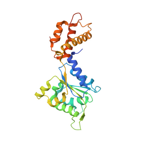
2BJV

Crystal Structure of PspF(1-275) R168A mutant


Experimental Data Snapshot

  • Method: X-RAY DIFFRACTION
  • Resolution: 1.70 Å
  • R-Value Free: 0.212 
  • R-Value Work: 0.172 
  • R-Value Observed: 0.172 

wwPDB Validation   3D Report Full Report


This is version 1.3 of the entry. See complete history


Literature

Structural Insights Into the Activity of Enhancer-Binding Proteins

Rappas, M.Schumacher, J.Beuron, F.Niwa, H.Bordes, P.Wigneshweraraj, S.Keetch, C.A.Robinson, C.V.Buck, M.Zhang, X.

(2005) Science 307: 1972

  • DOI: https://doi.org/10.1126/science.1105932
  • Primary Citation of Related Structures:  
    2BJV, 2BJW

  • PubMed Abstract: 

    Activators of bacterial sigma54-RNA polymerase holoenzyme are mechanochemical proteins that use adenosine triphosphate (ATP) hydrolysis to activate transcription. We have determined by cryogenic electron microscopy (cryo-EM) a 20 angstrom resolution structure of an activator, phage shock protein F [PspF(1-275)], which is bound to an ATP transition state analog in complex with its basal factor, sigma54. By fitting the crystal structure of PspF(1-275) at 1.75 angstroms into the EM map, we identified two loops involved in binding sigma54. Comparing enhancer-binding structures in different nucleotide states and mutational analysis led us to propose nucleotide-dependent conformational changes that free the loops for association with sigma54.


  • Organizational Affiliation

    Department of Biological Sciences, Imperial College London, London, SW7 2AZ, UK.


Macromolecules
Find similar proteins by:  (by identity cutoff)  |  3D Structure
Entity ID: 1
MoleculeChains Sequence LengthOrganismDetailsImage
PSP OPERON TRANSCRIPTIONAL ACTIVATOR265Escherichia coliMutation(s): 1 
UniProt
Find proteins for P37344 (Escherichia coli (strain K12))
Explore P37344 
Go to UniProtKB:  P37344
Entity Groups  
Sequence Clusters30% Identity50% Identity70% Identity90% Identity95% Identity100% Identity
UniProt GroupP37344
Sequence Annotations
Expand
  • Reference Sequence
Experimental Data & Validation

Experimental Data

  • Method: X-RAY DIFFRACTION
  • Resolution: 1.70 Å
  • R-Value Free: 0.212 
  • R-Value Work: 0.172 
  • R-Value Observed: 0.172 
  • Space Group: P 65
Unit Cell:
Length ( Å )Angle ( ˚ )
a = 113.613α = 90
b = 113.613β = 90
c = 39.145γ = 120
Software Package:
Software NamePurpose
CNSrefinement
DENZOdata reduction
SCALEPACKdata scaling
SHARPphasing

Structure Validation

View Full Validation Report


Entry History 

Deposition Data

Revision History  (Full details and data files)

  • Version 1.0: 2005-03-31
    Type: Initial release
  • Version 1.1: 2011-05-08
    Changes: Version format compliance
  • Version 1.2: 2011-07-13
    Changes: Version format compliance
  • Version 1.3: 2017-08-30
    Changes: Data collection