2BJO | pdb_00002bjo

Crystal Structure of the Organic Hydroperoxide Resistance Protein OhrB of Bacillus subtilis


Experimental Data Snapshot

  • Method: X-RAY DIFFRACTION
  • Resolution: 2.10 Å
  • R-Value Free: 
    0.199 (Depositor), 0.204 (DCC) 
  • R-Value Work: 
    0.158 (Depositor), 0.170 (DCC) 
  • R-Value Observed: 
    0.161 (Depositor) 

Starting Model: experimental
View more details

wwPDB Validation 3D Report Full Report

Validation slider image for 2BJO

This is version 1.3 of the entry. See complete history

Literature

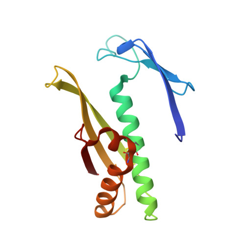
Structure of the Bacillus Subtilis Ohrb Hydroperoxide-Resistance Protein in a Fully Oxidized State.

Cooper, D.R.Surendranath, Y.Devedjiev, Y.Bielnicki, J.Derewenda, Z.S.

(2007) Acta Crystallogr D Biol Crystallogr 63: 1269

  • DOI: https://doi.org/10.1107/S0907444907050226
  • Primary Citation Related Structures: 
    2BJO

  • PubMed Abstract: 

    The crystal structure of the fully oxidized form of the Bacillus subtilis organic hydroperoxide-resistance (OhrB) protein is reported at 2.1 A resolution. The electron density reveals an intact catalytic disulfide bond (Cys55-Cys119) in each of the two molecules, which are intertwined into a canonical obligate dimer. However, the stereochemistry of the disulfides is unorthodox and strained, suggesting that they are sensitive to reducing agents. A deep solvent-accessible gorge reaching Cys55 may represent the access route for the reductant.


  • Organizational Affiliation
    • Department of Molecular Physiology and Biological Physics, University of Virginia and the PSI2 Integrated Center for Structure-Function Innovation Charlottesville, Virginia 22908, USA.

Macromolecule Content 

  • Total Structure Weight: 28.89 kDa 
  • Atom Count: 2,224 
  • Modeled Residue Count: 270 
  • Deposited Residue Count: 272 
  • Unique protein chains: 1

Macromolecules

Find similar proteins by:|  3D Structure
Entity ID: 1
MoleculeChains  Sequence LengthOrganismDetailsImage
ORGANIC HYDROPEROXIDE RESISTANCE PROTEIN OHRB
A, B
136Bacillus subtilisMutation(s): 3 
UniProt
Find proteins for P80242 (Bacillus subtilis (strain 168))
Explore P80242 
Go to UniProtKB:  P80242
Entity Groups
Sequence Clusters30% Identity50% Identity70% Identity90% Identity95% Identity100% Identity
UniProt GroupP80242
Sequence Annotations
Expand
Reference Sequence

Experimental Data & Validation

Experimental Data

  • Method: X-RAY DIFFRACTION
  • Resolution: 2.10 Å
  • R-Value Free:  0.199 (Depositor), 0.204 (DCC) 
  • R-Value Work:  0.158 (Depositor), 0.170 (DCC) 
  • R-Value Observed: 0.161 (Depositor) 
Space Group: P 1
Unit Cell:
Length ( Å )Angle ( ˚ )
a = 35.34α = 84.45
b = 41.434β = 91.51
c = 44.6γ = 73.89
Software Package:
Software NamePurpose
REFMACrefinement
HKL-2000data reduction
HKL-2000data scaling
AMoREphasing

Structure Validation

View Full Validation Report


Entry History 

Deposition Data

Revision History  (Full details and data files)

  • Version 1.0: 2006-03-09
    Type: Initial release
  • Version 1.1: 2011-07-13
    Changes: Advisory, Version format compliance
  • Version 1.2: 2023-12-13
    Changes: Data collection, Database references, Other, Refinement description
  • Version 1.3: 2024-11-13
    Changes: Structure summary