2BE4

X-RAY STRUCTURE AN EF-HAND PROTEIN FROM DANIO RERIO Dr.36843


Experimental Data Snapshot

  • Method: X-RAY DIFFRACTION
  • Resolution: 2.10 Å
  • R-Value Free: 0.253 
  • R-Value Work: 0.175 
  • R-Value Observed: 0.179 

wwPDB Validation   3D Report Full Report


This is version 1.4 of the entry. See complete history

Re-refinement Note

A newer entry is available that reflects an alternative modeling of the original data: 2Q4U


Literature

X-ray structure of Danio rerio secretagogin: A hexa-EF-hand calcium sensor.

Bitto, E.Bingman, C.A.Bittova, L.Frederick, R.O.Fox, B.G.Phillips Jr., G.N.

(2009) Proteins 76: 477-483

  • DOI: https://doi.org/10.1002/prot.22362

  • PubMed Abstract: 

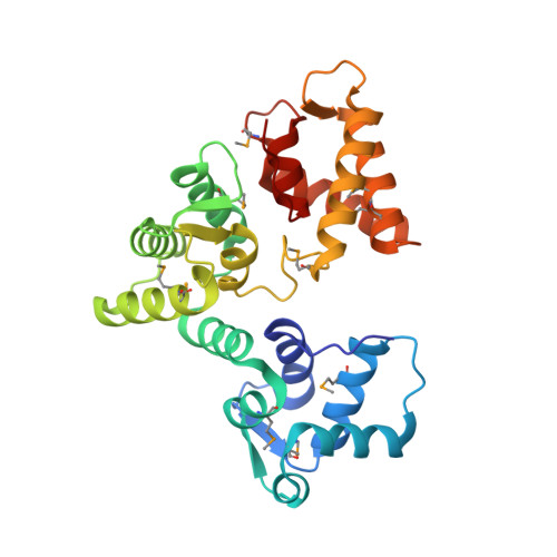
    Many essential physiological processes are regulated by the modulation of calcium concentration in the cell. The EF-hand proteins represent a superfamily of calcium-binding proteins involved in calcium signaling and homeostasis. Secretagogin is a hexa-EF-hand protein that is highly expressed in pancreatic islet of Langerhans and neuroendocrine cells and may play a role in the trafficking of secretory granules. We present the X-ray structure of Danio rerio secretagogin, which is 73% identical to human secretagogin, in calcium-free form at 2.1-A resolution. Secretagogin consists of the three globular domains each of which contains a pair of EF-hand motifs. The domains are arranged into a V-shaped molecule with a distinct groove formed at the interface of the domains. Comparison of the secretagogin structure with the solution structure of calcium-loaded calbindin D(28K) revealed a striking difference in the spatial arrangement of their domains, which involves approximately 180 degrees rotation of the first globular domain with respect to the module formed by the remaining domains.


  • Organizational Affiliation

    Department of Biochemistry, Center for Eukaryotic Structural Genomics, University of Wisconsin-Madison, 53706-1544, USA.


Macromolecules
Find similar proteins by:  (by identity cutoff)  |  3D Structure
Entity ID: 1
MoleculeChains Sequence LengthOrganismDetailsImage
hypothetical protein LOC449832272Danio rerioMutation(s): 10 
Gene Names: Dr.36843BC083168
UniProt
Find proteins for Q5XJX1 (Danio rerio)
Explore Q5XJX1 
Go to UniProtKB:  Q5XJX1
Entity Groups  
Sequence Clusters30% Identity50% Identity70% Identity90% Identity95% Identity100% Identity
UniProt GroupQ5XJX1
Sequence Annotations
Expand
  • Reference Sequence
Small Molecules
Modified Residues  1 Unique
IDChains TypeFormula2D DiagramParent
MSE
Query on MSE
A
L-PEPTIDE LINKINGC5 H11 N O2 SeMET
Experimental Data & Validation

Experimental Data

  • Method: X-RAY DIFFRACTION
  • Resolution: 2.10 Å
  • R-Value Free: 0.253 
  • R-Value Work: 0.175 
  • R-Value Observed: 0.179 
  • Space Group: P 21 21 21
Unit Cell:
Length ( Å )Angle ( ˚ )
a = 47.884α = 90
b = 52.748β = 90
c = 114.364γ = 90
Software Package:
Software NamePurpose
DENZOdata reduction
SCALEPACKdata scaling
SHARPphasing
DMphasing
REFMACrefinement
PDB_EXTRACTdata extraction
PHENIXphasing
SHELXDphasing
SOLOMONphasing
ARP/wARPmodel building

Structure Validation

View Full Validation Report


Entry History 

Revision History  (Full details and data files)

  • Version 1.0: 2005-11-01
    Type: Initial release
  • Version 1.1: 2008-05-01
    Changes: Version format compliance
  • Version 1.2: 2011-07-13
    Changes: Advisory, Version format compliance
  • Version 1.3: 2013-02-06
    Changes: Database references
  • Version 1.4: 2017-10-18
    Changes: Refinement description