2BCW

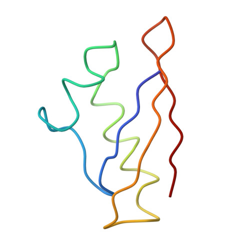
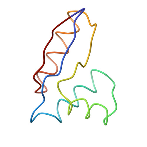
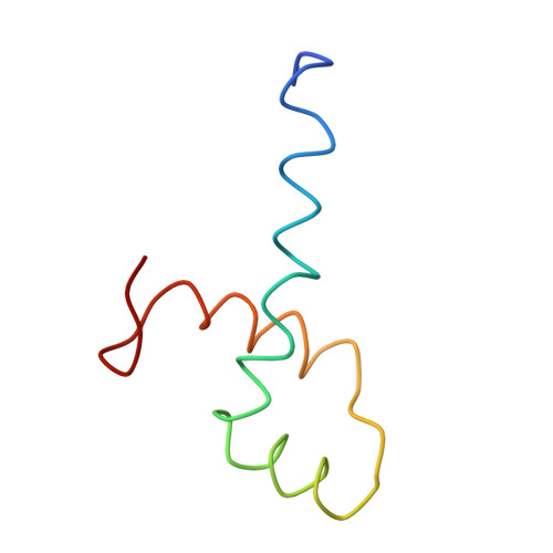
Coordinates of the N-terminal domain of ribosomal protein L11,C-terminal domain of ribosomal protein L7/L12 and a portion of the G' domain of elongation factor G, as fitted into cryo-em map of an Escherichia coli 70S*EF-G*GDP*fusidic acid complex


Experimental Data Snapshot

  • Method: ELECTRON MICROSCOPY
  • Resolution: 11.2 Å
  • Aggregation State: PARTICLE 
  • Reconstruction Method: SINGLE PARTICLE 

wwPDB Validation   3D Report Full Report


This is version 1.3 of the entry. See complete history


Literature

Interaction of the G' Domain of Elongation Factor G and the C-Terminal Domain of Ribosomal Protein L7/L12 during Translocation as Revealed by Cryo-EM.

Datta, P.P.Sharma, M.R.Qi, L.Frank, J.Agrawal, R.K.

(2005) Mol Cell 20: 723-731

  • DOI: https://doi.org/10.1016/j.molcel.2005.10.028
  • Primary Citation of Related Structures:  
    2BCW

  • PubMed Abstract: 
  • During tRNA translocation on the ribosome, an arc-like connection (ALC) is formed between the G' domain of elongation factor G (EF-G) and the L7/L12-stalk base of the large ribosomal subunit in the GDP state. To delineate the boundary of EF-G within the ALC, we tagged an amino acid residue near the tip of the G' domain of EF-G with undecagold, which was then visualized with three-dimensional cryo-electron microscopy (cryo-EM) ...

    During tRNA translocation on the ribosome, an arc-like connection (ALC) is formed between the G' domain of elongation factor G (EF-G) and the L7/L12-stalk base of the large ribosomal subunit in the GDP state. To delineate the boundary of EF-G within the ALC, we tagged an amino acid residue near the tip of the G' domain of EF-G with undecagold, which was then visualized with three-dimensional cryo-electron microscopy (cryo-EM). Two distinct positions for the undecagold, observed in the GTP-state and GDP-state cryo-EM maps of the ribosome bound EF-G, allowed us to determine the movement of the labeled amino acid. Molecular analyses of the cryo-EM maps show: (1) that three structural components, the N-terminal domain of ribosomal protein L11, the C-terminal domain of ribosomal protein L7/L12, and the G' domain of EF-G, participate in formation of the ALC; and (2) that both EF-G and the ribosomal protein L7/L12 undergo large conformational changes to form the ALC.


    Related Citations: 
    • A detailed view of a ribosomal active site: the structure of the L11-RNA complex
      Wimberly, B.T., Guymon, R., McCutcheon, J.P., White, S.W., Ramakrishnan, V.
      (1999) Cell 97: 491
    • Structure of the C-terminal domain of the ribosomal protein L7/L12 from Escherichia coli at 1.7
      Leijonmarck, M., Liljas, A.
      (1987) J Mol Biol 195: 555
    • Three-dimensional structure of the ribosomal translocase: elongation factor G from Thermus thermophilus
      Aevarsson, A., Brazhnikov, E., Garber, M., Zheltonosova, J., Chirgadze, Y., al-Karadaghi, S., Svensson, L.A., Liljas, A.
      (1994) EMBO J 13: 3669

    Organizational Affiliation

    Division of Molecular Medicine, Wadsworth Center, New York State Department of Health, Empire State Plaza, P.O. Box 509, Albany, New York 12201, USA.


Macromolecules
Find similar proteins by:  (by identity cutoff)  |  3D Structure
Entity ID: 1
MoleculeChainsSequence LengthOrganismDetailsImage
50S ribosomal protein L1165Thermotoga maritimaMutation(s): 0 
UniProt
Find proteins for P29395 (Thermotoga maritima (strain ATCC 43589 / DSM 3109 / JCM 10099 / NBRC 100826 / MSB8))
Explore P29395 
Go to UniProtKB:  P29395
Entity Groups  
Sequence Clusters30% Identity50% Identity70% Identity90% Identity95% Identity100% Identity
UniProt GroupP29395
Protein Feature View
Expand
  • Reference Sequence
Find similar proteins by:  (by identity cutoff)  |  3D Structure
Entity ID: 2
MoleculeChainsSequence LengthOrganismDetailsImage
50S ribosomal protein L7/L1268Escherichia coliMutation(s): 0 
UniProt
Find proteins for P0A7K2 (Escherichia coli (strain K12))
Explore P0A7K2 
Go to UniProtKB:  P0A7K2
Entity Groups  
Sequence Clusters30% Identity50% Identity70% Identity90% Identity95% Identity100% Identity
UniProt GroupP0A7K2
Protein Feature View
Expand
  • Reference Sequence
Find similar proteins by:  (by identity cutoff)  |  3D Structure
Entity ID: 3
MoleculeChainsSequence LengthOrganismDetailsImage
Elongation factor G58Thermus thermophilusMutation(s): 0 
Gene Names: fusAfus
UniProt
Find proteins for P13551 (Thermus thermophilus)
Explore P13551 
Go to UniProtKB:  P13551
Entity Groups  
Sequence Clusters30% Identity50% Identity70% Identity90% Identity95% Identity100% Identity
UniProt GroupP13551
Protein Feature View
Expand
  • Reference Sequence
Experimental Data & Validation

Experimental Data

  • Method: ELECTRON MICROSCOPY
  • Resolution: 11.2 Å
  • Aggregation State: PARTICLE 
  • Reconstruction Method: SINGLE PARTICLE 
EM Software:
TaskSoftware PackageVersion
RECONSTRUCTIONSPIDER

Structure Validation

View Full Validation Report


Entry History 

Deposition Data

Revision History  (Full details and data files)

  • Version 1.0: 2005-12-20
    Type: Initial release
  • Version 1.1: 2008-05-01
    Changes: Version format compliance
  • Version 1.2: 2011-07-13
    Changes: Version format compliance
  • Version 1.3: 2018-07-18
    Changes: Data collection