2B1X

Crystal structure of naphthalene 1,2-dioxygenase from Rhodococcus sp.


Experimental Data Snapshot

  • Method: X-RAY DIFFRACTION
  • Resolution: 2.00 Å
  • R-Value Free: 0.224 
  • R-Value Work: 0.191 
  • R-Value Observed: 0.191 

wwPDB Validation   3D Report Full Report


This is version 1.3 of the entry. See complete history


Literature

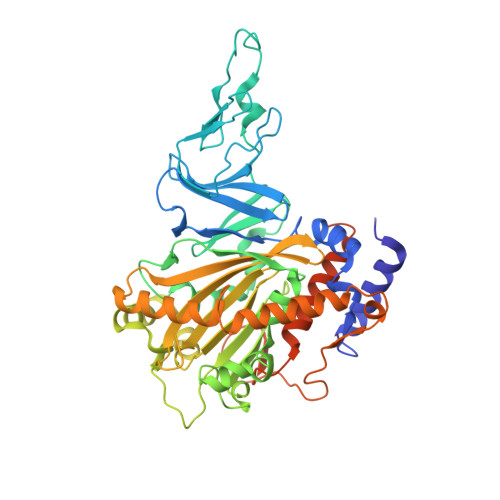
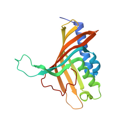
Structure and Increased Thermostability of Rhodococcus sp. Naphthalene 1,2-Dioxygenase.

Gakhar, L.Malik, Z.A.Allen, C.C.Lipscomb, D.A.Larkin, M.J.Ramaswamy, S.

(2005) J Bacteriol 187: 7222-7231

  • DOI: https://doi.org/10.1128/JB.187.21.7222-7231.2005
  • Primary Citation of Related Structures:  
    2B1X, 2B24

  • PubMed Abstract: 

    Rieske nonheme iron oxygenases form a large class of aromatic ring-hydroxylating dioxygenases found in microorganisms. These enzymes enable microorganisms to tolerate and even exclusively utilize aromatic compounds for growth, making them good candidates for use in synthesis of chiral intermediates and bioremediation. Studies of the chemical stability and thermostability of these enzymes thus become important. We report here the structure of free and substrate (indole)-bound forms of naphthalene dioxygenase from Rhodococcus sp. strain NCIMB12038. The structure of the Rhodococcus enzyme reveals that, despite a approximately 30% sequence identity between these naphthalene dioxygenases, their overall structures superpose very well with a root mean square deviation of less than 1.6 A. The differences in the active site of the two enzymes are pronounced near the entrance; however, indole binds to the Rhodococcus enzyme in the same orientation as in the Pseudomonas enzyme. Circular dichroism spectroscopy experiments show that the Rhodococcus enzyme has higher thermostability than the naphthalene dioxygenase from Pseudomonas species. The Pseudomonas enzyme has an apparent melting temperature of 55 degrees C while the Rhodococcus enzyme does not completely unfold even at 95 degrees C. Both enzymes, however, show similar unfolding behavior in urea, and the Rhodococcus enzyme is only slightly more tolerant to unfolding by guanidine hydrochloride. Structure analysis suggests that the higher thermostability of the Rhodococcus enzyme may be attributed to a larger buried surface area and extra salt bridge networks between the alpha and beta subunits in the Rhodococcus enzyme.


  • Organizational Affiliation

    Department of Biochemistry, University of Iowa, Iowa City, IA 52242, USA.


Macromolecules
Find similar proteins by:  (by identity cutoff)  |  3D Structure
Entity ID: 1
MoleculeChains Sequence LengthOrganismDetailsImage
naphthalene dioxygenase large subunit
A, C, E
470Rhodococcus sp. NCIMB 12038Mutation(s): 0 
EC: 1.14.12.12
UniProt
Find proteins for Q9X3R9 (Rhodococcus sp. NCIMB 12038)
Explore Q9X3R9 
Go to UniProtKB:  Q9X3R9
Entity Groups  
Sequence Clusters30% Identity50% Identity70% Identity90% Identity95% Identity100% Identity
UniProt GroupQ9X3R9
Sequence Annotations
Expand
  • Reference Sequence
Find similar proteins by:  (by identity cutoff)  |  3D Structure
Entity ID: 2
MoleculeChains Sequence LengthOrganismDetailsImage
naphthalene dioxygenase small subunit
B, D, F
172Rhodococcus sp. NCIMB 12038Mutation(s): 0 
EC: 1.14.12.12
UniProt
Find proteins for Q7BSK6 (Rhodococcus sp. NCIMB 12038)
Explore Q7BSK6 
Go to UniProtKB:  Q7BSK6
Entity Groups  
Sequence Clusters30% Identity50% Identity70% Identity90% Identity95% Identity100% Identity
UniProt GroupQ7BSK6
Sequence Annotations
Expand
  • Reference Sequence
Small Molecules
Ligands 3 Unique
IDChains Name / Formula / InChI Key2D Diagram3D Interactions
FES
Query on FES

Download Ideal Coordinates CCD File 
H [auth A],
N [auth C],
S [auth E]
FE2/S2 (INORGANIC) CLUSTER
Fe2 S2
NIXDOXVAJZFRNF-UHFFFAOYSA-N
MPD
Query on MPD

Download Ideal Coordinates CCD File 
I [auth A]
J [auth A]
K [auth A]
L [auth A]
O [auth C]
I [auth A],
J [auth A],
K [auth A],
L [auth A],
O [auth C],
P [auth C],
Q [auth C],
T [auth E],
U [auth E],
V [auth E],
W [auth E],
X [auth E],
Y [auth E]
(4S)-2-METHYL-2,4-PENTANEDIOL
C6 H14 O2
SVTBMSDMJJWYQN-YFKPBYRVSA-N
FE
Query on FE

Download Ideal Coordinates CCD File 
G [auth A],
M [auth C],
R [auth E]
FE (III) ION
Fe
VTLYFUHAOXGGBS-UHFFFAOYSA-N
Experimental Data & Validation

Experimental Data

  • Method: X-RAY DIFFRACTION
  • Resolution: 2.00 Å
  • R-Value Free: 0.224 
  • R-Value Work: 0.191 
  • R-Value Observed: 0.191 
  • Space Group: P 21 21 21
Unit Cell:
Length ( Å )Angle ( ˚ )
a = 87.744α = 90
b = 144.246β = 90
c = 186.207γ = 90
Software Package:
Software NamePurpose
REFMACrefinement
SBC-Collectdata collection
d*TREKdata scaling
AMoREphasing

Structure Validation

View Full Validation Report


Entry History 

Deposition Data

Revision History  (Full details and data files)

  • Version 1.0: 2005-10-04
    Type: Initial release
  • Version 1.1: 2008-05-01
    Changes: Version format compliance
  • Version 1.2: 2011-07-13
    Changes: Advisory, Source and taxonomy, Version format compliance
  • Version 1.3: 2023-08-23
    Changes: Data collection, Database references, Derived calculations, Refinement description