2AYG

Crystal structure of HPV6a E2 DNA binding domain bound to an 18 base pair DNA target


Experimental Data Snapshot

  • Method: X-RAY DIFFRACTION
  • Resolution: 3.10 Å
  • R-Value Free: 0.288 
  • R-Value Work: 0.203 
  • R-Value Observed: 0.247 

wwPDB Validation   3D Report Full Report


This is version 1.3 of the entry. See complete history


Literature

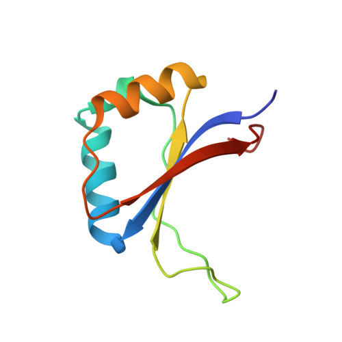
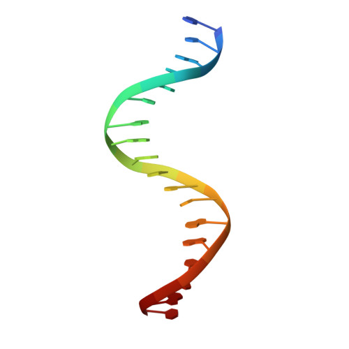
The recognition of local DNA conformation by the human papillomavirus type 6 E2 protein.

Hooley, E.Fairweather, V.Clarke, A.R.Gaston, K.Brady, R.L.

(2006) Nucleic Acids Res 34: 3897-3908

  • DOI: https://doi.org/10.1093/nar/gkl466
  • Primary Citation of Related Structures:  
    2AYB, 2AYE, 2AYG

  • PubMed Abstract: 

    The E2 proteins are transcription/replication factors from papillomaviruses. Human papillomaviruses (HPVs) can be broadly divided in two groups; low-risk HPV subtypes cause benign warts while high-risk HPVs give rise to cervical cancer. Although a range of crystal structures of E2 DNA-binding domains (DBD) from both high- and low-risk HPV subtypes have been reported previously, structures of E2 DBD:DNA complexes have only been available for high-risk HPV18 and bovine papillomavirus (BPV1). In the present study we report the unliganded and DNA complex structures of the E2 DBD from the low-risk HPV6. As in the previous E2-DNA structures, complex formation results in considerable bending of the DNA, which is facilitated by sequences with A:T-rich spacers that adopt a pre-bent conformation. The low-risk HPV6 E2-DNA complex differs from the earlier structures in that minimal deformation of the protein accompanies complex formation. Stopped-flow kinetic studies confirm that both high- and low-risk E2 proteins adapt their structures on binding to DNA, although this is achieved more readily for HPV6 E2. It therefore appears that the higher selectivity of the HPV6 E2 protein may arise from its limited molecular adaptability, a property that might distinguish the behaviour of E2 proteins from high- and low-risk HPV subtypes.


  • Organizational Affiliation

    Department of Biochemistry, University of Bristol, Bristol BS8 1TD, UK.


Macromolecules

Find similar proteins by:  (by identity cutoff)  |  3D Structure
Entity ID: 2
MoleculeChains Sequence LengthOrganismDetailsImage
Regulatory protein E2C [auth A],
D [auth B]
87Human papillomavirus type 6aMutation(s): 1 
Gene Names: E2
UniProt
Find proteins for Q84294 (Human papillomavirus type 6a)
Explore Q84294 
Go to UniProtKB:  Q84294
Entity Groups  
Sequence Clusters30% Identity50% Identity70% Identity90% Identity95% Identity100% Identity
UniProt GroupQ84294
Sequence Annotations
Expand
  • Reference Sequence

Find similar nucleic acids by:  Sequence   |   3D Structure  

Entity ID: 1
MoleculeChains LengthOrganismImage
5'-D(*GP*CP*AP*AP*CP*CP*GP*AP*AP*TP*TP*CP*GP*GP*TP*TP*GP*C)-3'A [auth C],
B [auth D]
18N/A
Sequence Annotations
Expand
  • Reference Sequence
Experimental Data & Validation

Experimental Data

  • Method: X-RAY DIFFRACTION
  • Resolution: 3.10 Å
  • R-Value Free: 0.288 
  • R-Value Work: 0.203 
  • R-Value Observed: 0.247 
  • Space Group: P 61
Unit Cell:
Length ( Å )Angle ( ˚ )
a = 73.449α = 90
b = 73.449β = 90
c = 109.237γ = 120
Software Package:
Software NamePurpose
PHASERphasing
REFMACrefinement
PDB_EXTRACTdata extraction
HKL-2000data reduction
SCALEPACKdata scaling

Structure Validation

View Full Validation Report


Entry History 

Deposition Data

Revision History  (Full details and data files)

  • Version 1.0: 2006-08-22
    Type: Initial release
  • Version 1.1: 2008-05-01
    Changes: Version format compliance
  • Version 1.2: 2011-07-13
    Changes: Version format compliance
  • Version 1.3: 2023-08-23
    Changes: Data collection, Database references, Refinement description