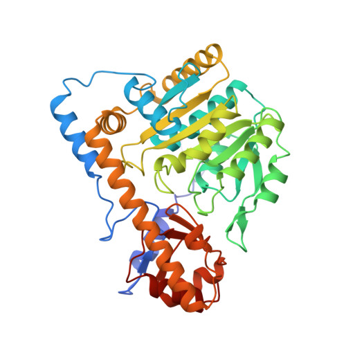
The active site of Paracoccus denitrificans aromatic amino acid aminotransferase has contrary properties: flexibility and rigidity.
Okamoto, A., Ishii, S., Hirotsu, K., Kagamiyama, H.(1999) Biochemistry 38: 1176-1184
- PubMed: 9930977
- DOI: https://doi.org/10.1021/bi981921d
- Primary Citation of Related Structures:
2AY1, 2AY2, 2AY3, 2AY4, 2AY5, 2AY6, 2AY7, 2AY8, 2AY9 - PubMed Abstract:
Paracoccus denitrificans aromatic amino acid aminotransferase (EC 2. 6.1.57; pdAroAT) binds with a series of aliphatic monocarboxylates attached to the bulky hydrophobic groups. To analyze the properties of the active site in this enzyme, we determined the tertiary structures of pdAroAT complexed with nine different inhibitors. Comparison of these active site structures showed that the active site of pdAroAT consists of two parts with contrary properties: rigidity and flexibility. The regions that interact with the carboxylates and methylene chains of the inhibitors gave essentially the same structures among these complexes, exhibiting the rigid property, which would involve fixing the substrate at the proper orientation for efficient catalysis. The region that interacts with the terminal hydrophobic groups of the inhibitors gave versatile structures according to the structures of the terminal groups, showing that this region is structurally flexible. This is mainly achieved by the conformational versatility of the side chains of Asp15, Lys16, Asn142, Arg292, and Ser296. These residues formed in the active site hydrogen bond networks, which were adaptable for the structures of the terminal hydrophobic groups of the inhibitors, with a small deformation or partial destruction according to the shapes and sizes of the inhibitors. These observations illustrate how the flexibility and rigidity in the active site can be used for the substrate binding and recognition.
- Department of Biochemistry, Osaka Medical College, Takatsuki, Osaka 569-8686, Japan.
Organizational Affiliation:


