2AT1

CRYSTAL STRUCTURES OF PHOSPHONOACETAMIDE LIGATED T AND PHOSPHONOACETAMIDE AND MALONATE LIGATED R STATES OF ASPARTATE CARBAMOYLTRANSFERASE AT 2.8-ANGSTROMS RESOLUTION AND NEUTRAL PH


Experimental Data Snapshot

  • Method: X-RAY DIFFRACTION
  • Resolution: 2.80 Å
  • R-Value Work: 0.170 

wwPDB Validation   3D Report Full Report


This is version 2.1 of the entry. See complete history


Literature

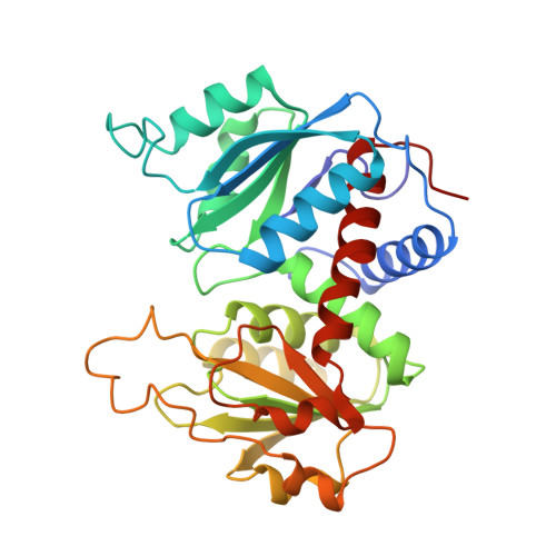
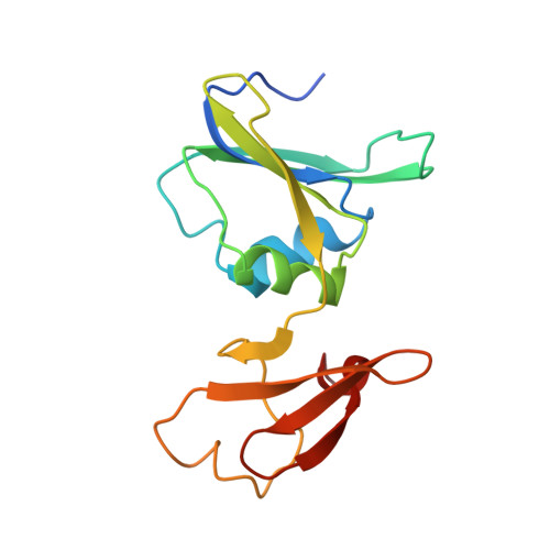
Crystal structures of phosphonoacetamide ligated T and phosphonoacetamide and malonate ligated R states of aspartate carbamoyltransferase at 2.8-A resolution and neutral pH.

Gouaux, J.E.Lipscomb, W.N.

(1990) Biochemistry 29: 389-402

  • DOI: https://doi.org/10.1021/bi00454a013
  • Primary Citation of Related Structures:  
    1AT1, 2AT1, 3AT1

  • PubMed Abstract: 

    The T----R transition of the cooperative enzyme aspartate carbamoyltransferase occurs at pH 7 in single crystals without visibly cracking many of the crystals and leaving those uncracked suitable for single-crystal X-ray analysis. To promote the T----R transition, we employ the competitive inhibitors of carbamoyl phosphate and aspartate, which are phosphonoacetamide (PAM) and malonate, respectively. In response to PAM binding to the T-state crystals, residues Thr 53-Thr 55 and Pro 266-Pro 268 move to their R-state positions to bind to the phosphonate and amino group of PAM. These changes induce a conformation that can bind tightly the aspartate analogue malonate, which thereby effects the allosteric transition. We prove this by showing that PAM-ligated T-state crystals (Tpam), space group P321 (a = 122.2 A, c = 142.2 A), when transferred to a solution containing 20 mM PAM and 8 mM malonate at pH 7, isomerize to R-state crystals (Rpam,mal,soak), space group also P321 (a = 122.2 A, c = 156.4 A). The R-state structure in which the T----R transition occurs within the crystal at pH 7 compares very well (rms = 0.19 A for all atoms) with an R-state structure determined at pH 7 in which the crystals were initially grown in a solution of PAM and malonate at pH 5.9 and subsequently transferred to a buffer containing the ligands at pH 7 (Rpam,mal,crys). In fact, both of the PAM and malonate ligated R-state structures are very similar to both the carbamoyl phosphate and succinate or the N-(phosphonoacetyl)-L-aspartate ligated structures, even though the R-state structures reported here were determined at pH 7. Crystallographic residuals refined to 0.16-0.18 at 2.8-A resolution for the three structures.


  • Organizational Affiliation

    Gibbs Chemical Laboratory, Harvard University, Cambridge, Massachusetts 02138.


Macromolecules
Find similar proteins by:  (by identity cutoff)  |  3D Structure
Entity ID: 1
MoleculeChains Sequence LengthOrganismDetailsImage
ASPARTATE CARBAMOYLTRANSFERASE (R STATE), CATALYTIC CHAIN
A, C
310Escherichia coliMutation(s): 0 
EC: 2.1.3.2
UniProt
Find proteins for P0A786 (Escherichia coli (strain K12))
Explore P0A786 
Go to UniProtKB:  P0A786
Entity Groups  
Sequence Clusters30% Identity50% Identity70% Identity90% Identity95% Identity100% Identity
UniProt GroupP0A786
Sequence Annotations
Expand
  • Reference Sequence
Find similar proteins by:  (by identity cutoff)  |  3D Structure
Entity ID: 2
MoleculeChains Sequence LengthOrganismDetailsImage
ASPARTATE CARBAMOYLTRANSFERASE REGULATORY CHAIN
B, D
153Escherichia coliMutation(s): 0 
UniProt
Find proteins for P0A7F3 (Escherichia coli (strain K12))
Explore P0A7F3 
Go to UniProtKB:  P0A7F3
Entity Groups  
Sequence Clusters30% Identity50% Identity70% Identity90% Identity95% Identity100% Identity
UniProt GroupP0A7F3
Sequence Annotations
Expand
  • Reference Sequence
Oligosaccharides

Help

Entity ID: 3
MoleculeChains Length2D Diagram Glycosylation3D Interactions
alpha-D-glucopyranose-(1-4)-alpha-D-glucopyranose
E, F
2N/A
Glycosylation Resources
GlyTouCan:  G07411ON
GlyCosmos:  G07411ON
Biologically Interesting Molecules (External Reference) 1 Unique
Experimental Data & Validation

Experimental Data

  • Method: X-RAY DIFFRACTION
  • Resolution: 2.80 Å
  • R-Value Work: 0.170 
  • Space Group: P 3 2 1
Unit Cell:
Length ( Å )Angle ( ˚ )
a = 122.2α = 90
b = 122.2β = 90
c = 156.6γ = 120
Software Package:
Software NamePurpose
X-PLORrefinement

Structure Validation

View Full Validation Report


Entry History 

Deposition Data

Revision History  (Full details and data files)

  • Version 1.0: 1990-10-15
    Type: Initial release
  • Version 1.1: 2008-03-03
    Changes: Version format compliance
  • Version 1.2: 2011-07-13
    Changes: Derived calculations, Version format compliance
  • Version 1.3: 2017-11-29
    Changes: Advisory, Derived calculations, Other
  • Version 2.0: 2020-07-29
    Type: Remediation
    Reason: Carbohydrate remediation
    Changes: Advisory, Atomic model, Data collection, Database references, Derived calculations, Non-polymer description, Structure summary
  • Version 2.1: 2024-02-14
    Changes: Data collection, Database references, Structure summary