2AQ9 | pdb_00002aq9

Structure of E. coli LpxA with a bound peptide that is competitive with acyl-ACP


Experimental Data Snapshot

  • Method: X-RAY DIFFRACTION
  • Resolution: 1.80 Å
  • R-Value Free: 
    0.226 (Depositor), 0.237 (DCC) 
  • R-Value Work: 
    0.194 (Depositor), 0.211 (DCC) 

Starting Model: experimental
View more details

wwPDB Validation 3D Report Full Report

Validation slider image for 2AQ9

This is version 1.3 of the entry. See complete history

Literature

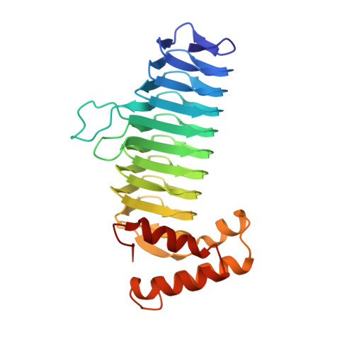
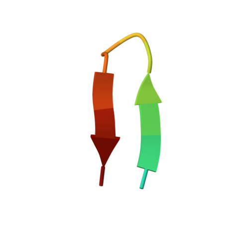
Structure of UDP-N-acetylglucosamine acyltransferase with a bound antibacterial pentadecapeptide.

Williams, A.H.Immormino, R.M.Gewirth, D.T.Raetz, C.R.

(2006) Proc Natl Acad Sci U S A 103: 10877-10882

  • DOI: https://doi.org/10.1073/pnas.0604465103
  • Primary Citation Related Structures: 
    2AQ9

  • PubMed Abstract: 

    UDP-GlcNAc acyltransferase (LpxA) catalyzes the first step of lipid A biosynthesis, the transfer of the R-3-hydroxyacyl chain from R-3-hydroxyacyl acyl carrier protein (ACP) to the glucosamine 3-OH group of UDP-GlcNAc. LpxA is essential for the growth of Escherichia coli and related Gram-negative bacteria. The crystal structure of the E. coli LpxA homotrimer, determined previously at 2.6 A in the absence of substrates or inhibitors, revealed that LpxA contains an unusual, left-handed parallel beta-helix fold. We now present the crystal structure at 1.8 A resolution of E. coli LpxA in a complex with a pentadecapeptide, peptide 920. Three peptides, each of which adopts a beta-hairpin conformation, are bound per LpxA trimer. The peptides are located at the interfaces of adjacent subunits in the vicinity of the three active sites. Each peptide interacts with residues from both adjacent subunits. Peptide 920 is a potent inhibitor of E. coli LpxA (Ki = 50 nM). It is competitive with respect to acyl-ACP but not UDP-GlcNAc. The compact beta-turn structure of peptide 920 bound to LpxA may open previously uncharacterized approaches to the rational design of LpxA inhibitors with antibiotic activity.


  • Organizational Affiliation
    • Department of Biochemistry, Duke University Medical Center, Box 3711 DUMC, Durham, NC 27710, USA.

Macromolecule Content 

  • Total Structure Weight: 32 kDa 
  • Atom Count: 2,567 
  • Modeled Residue Count: 274 
  • Deposited Residue Count: 277 
  • Unique protein chains: 2

Macromolecules

Find similar proteins by:|  3D Structure
Entity ID: 1
MoleculeChains  Sequence LengthOrganismDetailsImage
Acyl-[acyl-carrier-protein]--UDP-N-acetylglucosamine O-acyltransferase262Escherichia coli K-12Mutation(s): 0 
Gene Names: lpxA
EC: 2.3.1.129
UniProt
Find proteins for P0A722 (Escherichia coli (strain K12))
Explore P0A722 
Go to UniProtKB:  P0A722
Entity Groups
Sequence Clusters30% Identity50% Identity70% Identity90% Identity95% Identity100% Identity
UniProt GroupP0A722
Sequence Annotations
Expand
Reference Sequence
Find similar proteins by:  Sequence   |   3D Structure  
Entity ID: 2
MoleculeChains  Sequence LengthOrganismDetailsImage
peptide inhibitorB [auth X]15N/AMutation(s): 0 

Small Molecules

Ligands 2 Unique
IDChains Name / Formula / InChI Key2D Diagram3D Interactions
PO4

Query on PO4


Download:Ideal Coordinates CCD File
C [auth A],
CA [auth X],
D [auth A],
E [auth A],
F [auth A]
PHOSPHATE ION
O4 P
NBIIXXVUZAFLBC-UHFFFAOYSA-K
DMS

Query on DMS


Download:Ideal Coordinates CCD File
AA [auth A]
BA [auth A]
G [auth A]
H [auth A]
I [auth A]
AA [auth A],
BA [auth A],
G [auth A],
H [auth A],
I [auth A],
J [auth A],
K [auth A],
L [auth A],
M [auth A],
N [auth A],
O [auth A],
P [auth A],
Q [auth A],
R [auth A],
S [auth A],
T [auth A],
U [auth A],
V [auth A],
W [auth A],
X [auth A],
Y [auth A],
Z [auth A]
DIMETHYL SULFOXIDE
C2 H6 O S
IAZDPXIOMUYVGZ-UHFFFAOYSA-N

Experimental Data & Validation

Experimental Data

  • Method: X-RAY DIFFRACTION
  • Resolution: 1.80 Å
  • R-Value Free:  0.226 (Depositor), 0.237 (DCC) 
  • R-Value Work:  0.194 (Depositor), 0.211 (DCC) 
Space Group: P 21 3
Unit Cell:
Length ( Å )Angle ( ˚ )
a = 96.734α = 90
b = 96.734β = 90
c = 96.734γ = 90
Software Package:
Software NamePurpose
CNSrefinement
DENZOdata reduction
SCALEPACKdata scaling
MOLREPphasing

Structure Validation

View Full Validation Report


Entry History 

Deposition Data

Revision History  (Full details and data files)

  • Version 1.0: 2006-06-27
    Type: Initial release
  • Version 1.1: 2008-04-30
    Changes: Version format compliance
  • Version 1.2: 2011-07-13
    Changes: Derived calculations, Source and taxonomy, Version format compliance
  • Version 1.3: 2023-08-23
    Changes: Data collection, Database references, Derived calculations, Refinement description