2AMO

Loose Dimer of a Bacillus subtilis Nitric Oxide Synthase


Experimental Data Snapshot

  • Method: X-RAY DIFFRACTION
  • Resolution: 2.60 Å
  • R-Value Free: 0.290 
  • R-Value Work: 0.274 
  • R-Value Observed: 0.276 

wwPDB Validation   3D Report Full Report


This is version 1.3 of the entry. See complete history


Literature

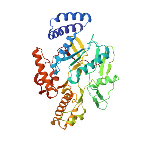
Structure of a loose dimer: an intermediate in nitric oxide synthase assembly.

Pant, K.Crane, B.R.

(2005) J Mol Biol 352: 932-940

  • DOI: https://doi.org/10.1016/j.jmb.2005.07.070
  • Primary Citation of Related Structures:  
    2AMO, 2AN0, 2AN2

  • PubMed Abstract: 

    Cooperativity among ligand binding, subunit association, and protein folding has implications for enzyme regulation as well as protein aggregation events associated with disease. The binding of substrate l-arginine or cofactor tetrahydrobiopterin converts nitric oxide synthases (NOSs) from a "loose dimer", with an exposed active center and higher sensitivity to proteolysis, to a "tight dimer" competent for catalysis. The crystallographic structure of the Bacillus subtilis NOS loose dimer shows an altered association state with severely destabilized subdomains. Ligand binding or heme reduction converts loose dimers to tight dimers in solution and crystals. Mutations at key positions in the dimer interface that distinguish prokaryotic from eukaryotic NOSs affect the propensity to form loose dimers. The loose dimer structure indicates that non-native interactions can mediate subunit association in NOS.


  • Organizational Affiliation

    Department of Chemistry and Chemical Biology, Cornell University Ithaca, NY 14853, USA.


Macromolecules
Find similar proteins by:  (by identity cutoff)  |  3D Structure
Entity ID: 1
MoleculeChains Sequence LengthOrganismDetailsImage
Nitric oxide synthase oxygenase
A, B
362Bacillus subtilisMutation(s): 0 
Gene Names: nos
EC: 1.14.13.39
UniProt
Find proteins for O34453 (Bacillus subtilis (strain 168))
Explore O34453 
Go to UniProtKB:  O34453
Entity Groups  
Sequence Clusters30% Identity50% Identity70% Identity90% Identity95% Identity100% Identity
UniProt GroupO34453
Sequence Annotations
Expand
  • Reference Sequence
Small Molecules
Ligands 1 Unique
IDChains Name / Formula / InChI Key2D Diagram3D Interactions
HEM
Query on HEM

Download Ideal Coordinates CCD File 
C [auth A],
D [auth B]
PROTOPORPHYRIN IX CONTAINING FE
C34 H32 Fe N4 O4
KABFMIBPWCXCRK-RGGAHWMASA-L
Experimental Data & Validation

Experimental Data

  • Method: X-RAY DIFFRACTION
  • Resolution: 2.60 Å
  • R-Value Free: 0.290 
  • R-Value Work: 0.274 
  • R-Value Observed: 0.276 
  • Space Group: P 21 21 2
Unit Cell:
Length ( Å )Angle ( ˚ )
a = 81.314α = 90
b = 93.211β = 90
c = 118.546γ = 90
Software Package:
Software NamePurpose
REFMACrefinement
HKL-2000data reduction
SCALEPACKdata scaling
AMoREphasing

Structure Validation

View Full Validation Report


Entry History 

Deposition Data

Revision History  (Full details and data files)

  • Version 1.0: 2006-10-31
    Type: Initial release
  • Version 1.1: 2008-04-30
    Changes: Version format compliance
  • Version 1.2: 2011-07-13
    Changes: Version format compliance
  • Version 1.3: 2023-08-23
    Changes: Data collection, Database references, Derived calculations, Refinement description