2ALD | pdb_00002ald

HUMAN MUSCLE ALDOLASE


Experimental Data Snapshot

  • Method: X-RAY DIFFRACTION
  • Resolution: 2.10 Å
  • R-Value Free: 
    0.258 (Depositor) 
  • R-Value Work: 
    0.172 (Depositor) 

wwPDB Validation 3D Report Full Report

Validation slider image for 2ALD

This is version 1.3 of the entry. See complete history

Literature

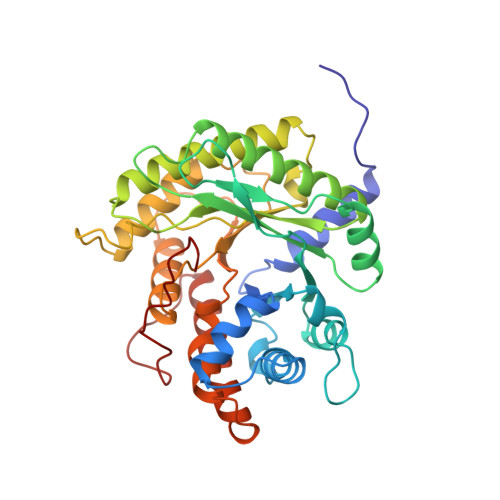
Crystal structure of human muscle aldolase complexed with fructose 1,6-bisphosphate: mechanistic implications.

Dalby, A.Dauter, Z.Littlechild, J.A.

(1999) Protein Sci 8: 291-297

  • DOI: https://doi.org/10.1110/ps.8.2.291
  • Primary Citation Related Structures: 
    2ALD, 4ALD

  • PubMed Abstract: 

    Fructose 1,6-bisphosphate aldolase catalyzes the reversible cleavage of fructose 1,6-bisphosphate and fructose 1-phosphate to dihydroxyacetone phosphate and either glyceraldehyde 3-phosphate or glyceraldehyde, respectively. Catalysis involves the formation of a Schiff's base intermediate formed at the epsilon-amino group of Lys229. The existing apo-enzyme structure was refined using the crystallographic free-R-factor and maximum likelihood methods that have been shown to give improved structural results that are less subject to model bias. Crystals were also soaked with the natural substrate (fructose 1,6-bisphosphate), and the crystal structure of this complex has been determined to 2.8 A. The apo structure differs from the previous Brookhaven-deposited structure (1ald) in the flexible C-terminal region. This is also the region where the native and complex structures exhibit differences. The conformational changes between native and complex structure are not large, but the observed complex does not involve the full formation of the Schiff's base intermediate, and suggests a preliminary hydrogen-bonded Michaelis complex before the formation of the covalent complex.


  • Organizational Affiliation
    • Department of Chemistry and Biological Sciences, Exeter University, United Kingdom.

Macromolecule Content 

  • Total Structure Weight: 39.34 kDa 
  • Atom Count: 2,906 
  • Modeled Residue Count: 363 
  • Deposited Residue Count: 363 
  • Unique protein chains: 1

Macromolecules

Find similar proteins by:|  3D Structure
Entity ID: 1
MoleculeChains  Sequence LengthOrganismDetailsImage
FRUCTOSE-BISPHOSPHATE ALDOLASE363Homo sapiensMutation(s): 0 
EC: 4.1.2.13
UniProt & NIH Common Fund Data Resources
Find proteins for P04075 (Homo sapiens)
Explore P04075 
Go to UniProtKB:  P04075
PHAROS:  P04075
GTEx:  ENSG00000149925 
Entity Groups
Sequence Clusters30% Identity50% Identity70% Identity90% Identity95% Identity100% Identity
UniProt GroupP04075
Sequence Annotations
Expand
Reference Sequence

Experimental Data & Validation

Experimental Data

  • Method: X-RAY DIFFRACTION
  • Resolution: 2.10 Å
  • R-Value Free:  0.258 (Depositor) 
  • R-Value Work:  0.172 (Depositor) 
Space Group: P 64 2 2
Unit Cell:
Length ( Å )Angle ( ˚ )
a = 96.5α = 90
b = 96.5β = 90
c = 167γ = 120
Software Package:
Software NamePurpose
AMoREphasing
REFMACrefinement
MOSFLMdata reduction
CCP4data scaling

Structure Validation

View Full Validation Report


Entry History 

Deposition Data

Revision History  (Full details and data files)

  • Version 1.0: 1999-04-20
    Type: Initial release
  • Version 1.1: 2008-03-24
    Changes: Version format compliance
  • Version 1.2: 2011-07-13
    Changes: Derived calculations, Version format compliance
  • Version 1.3: 2024-02-14
    Changes: Data collection, Database references