2AIR

T-state Active Site of Aspartate Transcarbamylase:Crystal Structure of the Carbamyl Phosphate and L-alanosine Ligated Enzyme


Experimental Data Snapshot

  • Method: X-RAY DIFFRACTION
  • Resolution: 2.00 Å
  • R-Value Free: 0.273 
  • R-Value Work: 0.239 
  • R-Value Observed: 0.239 

wwPDB Validation   3D Report Full Report


This is version 1.4 of the entry. See complete history


Literature

T-State Active Site of Aspartate Transcarbamylase: Crystal Structure of the Carbamyl Phosphate and l-Alanosine Ligated Enzyme

Huang, J.Lipscomb, W.N.

(2006) Biochemistry 45: 346-352

  • DOI: https://doi.org/10.1021/bi051543u
  • Primary Citation of Related Structures:  
    2AIR

  • PubMed Abstract: 

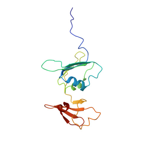
    An X-ray diffraction study to 2.0 A resolution shows that this enzyme, ATCase, is in the T-state (the c3 to c3 distance is 45.2 A) when ATCase is bound to carbamyl phosphate (CP) and to L-alanosine (an analogue of aspartate). This result strongly supports the kinetic results that alanosine did not inhibit the carbamylation of aspartate in the normal reaction of native ATCase plus CP and aspartate [Baillon, J., Tauc, P., and Hervé, G. (1985) Biochemistry 24, 7182-7187]. The structure further reveals that the phosphate of CP is 4 A away from its known position in the R-state and is in the T-state position of P(i) in a recent study of ATCase complexed with products, phosphate (P(i)) and N-carbamyl-L-aspartate [Huang, J., and Lipscomb, W. N. (2004) Biochemistry 43, 6422-6426]. Moreover, the alanosine position in this T-state is somewhat displaced from that expected for its analogue, aspartate, from the R-state position. The relations of these structural aspects to the kinetics are presented.


  • Organizational Affiliation

    Department of Chemistry and Chemical Biology, Harvard University, Cambridge, Massachusetts 02138, USA.


Macromolecules
Find similar proteins by:  (by identity cutoff)  |  3D Structure
Entity ID: 1
MoleculeChains Sequence LengthOrganismDetailsImage
Aspartate carbamoyltransferase catalytic chainA,
C [auth G]
310Escherichia coliMutation(s): 0 
Gene Names: pyrB
EC: 2.1.3.2
UniProt
Find proteins for P0A786 (Escherichia coli (strain K12))
Explore P0A786 
Go to UniProtKB:  P0A786
Entity Groups  
Sequence Clusters30% Identity50% Identity70% Identity90% Identity95% Identity100% Identity
UniProt GroupP0A786
Sequence Annotations
Expand
  • Reference Sequence
Find similar proteins by:  (by identity cutoff)  |  3D Structure
Entity ID: 2
MoleculeChains Sequence LengthOrganismDetailsImage
Aspartate carbamoyltransferase regulatory chainB,
D [auth H]
153Escherichia coliMutation(s): 0 
Gene Names: pyrI
UniProt
Find proteins for P0A7F3 (Escherichia coli (strain K12))
Explore P0A7F3 
Go to UniProtKB:  P0A7F3
Entity Groups  
Sequence Clusters30% Identity50% Identity70% Identity90% Identity95% Identity100% Identity
UniProt GroupP0A7F3
Sequence Annotations
Expand
  • Reference Sequence
Experimental Data & Validation

Experimental Data

  • Method: X-RAY DIFFRACTION
  • Resolution: 2.00 Å
  • R-Value Free: 0.273 
  • R-Value Work: 0.239 
  • R-Value Observed: 0.239 
  • Space Group: H 3
Unit Cell:
Length ( Å )Angle ( ˚ )
a = 129.152α = 90
b = 129.152β = 90
c = 198.097γ = 120
Software Package:
Software NamePurpose
HKL-2000data collection
SCALEPACKdata scaling
AMoREphasing
CNSrefinement
HKL-2000data reduction

Structure Validation

View Full Validation Report


Entry History 

Deposition Data

Revision History  (Full details and data files)

  • Version 1.0: 2006-01-24
    Type: Initial release
  • Version 1.1: 2008-04-30
    Changes: Version format compliance
  • Version 1.2: 2011-07-13
    Changes: Derived calculations, Version format compliance
  • Version 1.3: 2019-08-21
    Changes: Advisory, Data collection, Derived calculations
  • Version 1.4: 2023-08-23
    Changes: Data collection, Database references, Derived calculations, Refinement description