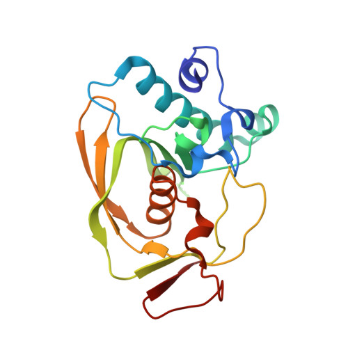
2AIE

S.pneumoniae polypeptide deformylase complexed with SB-505684


Experimental Data Snapshot

  • Method: X-RAY DIFFRACTION
  • Resolution: 1.70 Å
  • R-Value Free: 0.258 
  • R-Value Work: 0.227 

wwPDB Validation   3D Report Full Report


Ligand Structure Quality Assessment 


This is version 1.3 of the entry. See complete history


Literature

Structural Variation and inhibitor binding in polypeptide deformylase from four different bacterial species

Smith, K.J.Petit, C.M.Aubart, K.Smyth, M.McManus, E.Jones, J.Fosberry, A.Lewis, C.Lonetto, M.Christensen, S.B.

(2003) Protein Sci 12: 349-360

  • DOI: https://doi.org/10.1110/ps.0229303
  • Primary Citation of Related Structures:  
    2AI7, 2AI8, 2AI9, 2AIA, 2AIE

  • PubMed Abstract: 

    Polypeptide deformylase (PDF) catalyzes the deformylation of polypeptide chains in bacteria. It is essential for bacterial cell viability and is a potential antibacterial drug target. Here, we report the crystal structures of polypeptide deformylase from four different species of bacteria: Streptococcus pneumoniae, Staphylococcus aureus, Haemophilus influenzae, and Escherichia coli. Comparison of these four structures reveals significant overall differences between the two Gram-negative species (E. coli and H. influenzae) and the two Gram-positive species (S. pneumoniae and S. aureus). Despite these differences and low overall sequence identity, the S1' pocket of PDF is well conserved among the four enzymes studied. We also describe the binding of nonpeptidic inhibitor molecules SB-485345, SB-543668, and SB-505684 to both S. pneumoniae and E. coli PDF. Comparison of these structures shows similar binding interactions with both Gram-negative and Gram-positive species. Understanding the similarities and subtle differences in active site structure between species will help to design broad-spectrum polypeptide deformylase inhibitor molecules.


  • Organizational Affiliation

    GlaxoSmithKline, Harlow, Essex CM19 5AW, UK. Kathrine_J_Smith@gsk.com


Macromolecules
Find similar proteins by:  (by identity cutoff)  |  3D Structure
Entity ID: 1
MoleculeChains Sequence LengthOrganismDetailsImage
Peptide deformylaseA [auth P]203Streptococcus pneumoniaeMutation(s): 0 
Gene Names: def
EC: 3.5.1.88
UniProt
Find proteins for Q8DP79 (Streptococcus pneumoniae (strain ATCC BAA-255 / R6))
Explore Q8DP79 
Go to UniProtKB:  Q8DP79
Entity Groups  
Sequence Clusters30% Identity50% Identity70% Identity90% Identity95% Identity100% Identity
UniProt GroupQ8DP79
Sequence Annotations
Expand
  • Reference Sequence
Small Molecules
Binding Affinity Annotations 
IDSourceBinding Affinity
SB9 PDBBind:  2AIE IC50: 2200 (nM) from 1 assay(s)
BindingDB:  2AIE IC50: 2200 (nM) from 1 assay(s)
Experimental Data & Validation

Experimental Data

  • Method: X-RAY DIFFRACTION
  • Resolution: 1.70 Å
  • R-Value Free: 0.258 
  • R-Value Work: 0.227 
  • Space Group: P 43
Unit Cell:
Length ( Å )Angle ( ˚ )
a = 49.672α = 90
b = 49.672β = 90
c = 92.161γ = 90
Software Package:
Software NamePurpose
CNSrefinement
PDB_EXTRACTdata extraction

Structure Validation

View Full Validation Report


Ligand Structure Quality Assessment 


Entry History 

Deposition Data

Revision History  (Full details and data files)

  • Version 1.0: 2005-09-06
    Type: Initial release
  • Version 1.1: 2008-04-30
    Changes: Version format compliance
  • Version 1.2: 2011-07-13
    Changes: Version format compliance
  • Version 1.3: 2024-02-14
    Changes: Data collection, Database references, Derived calculations