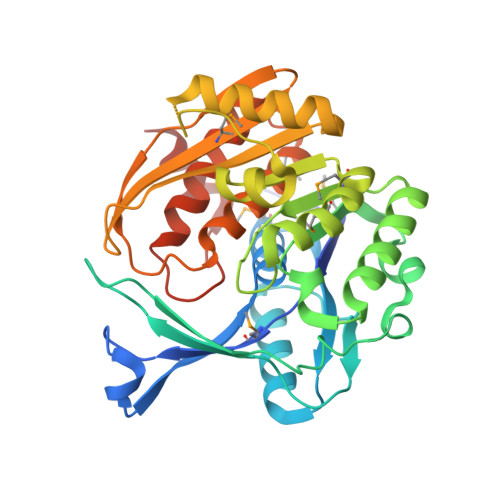
2AFB | pdb_00002afb

Crystal structure of 2-dehydro-3- deoxygluconokinase (EC 2.7.1.45) (tm0067) from THERMOTOGA MARITIMA at 2.05 A resolution


Experimental Data Snapshot

  • Method: X-RAY DIFFRACTION
  • Resolution: 2.05 Å
  • R-Value Free: 
    0.230 (Depositor), 0.239 (DCC) 
  • R-Value Work: 
    0.176 (Depositor), 0.187 (DCC) 
  • R-Value Observed: 
    0.177 (Depositor) 

wwPDB Validation 3D Report Full Report

Validation slider image for 2AFB

This is version 1.5 of the entry. See complete history. 

Literature

Crystal structure of 2-keto-3-deoxygluconate kinase (TM0067) from Thermotoga maritima at 2.05 A resolution.

Mathews, I.I., McMullan, D., Miller, M.D., Canaves, J.M., Elsliger, M.A., Floyd, R., Grzechnik, S.K., Jaroszewski, L., Klock, H.E., Koesema, E., Kovarik, J.S., Kreusch, A., Kuhn, P., McPhillips, T.M., Morse, A.T., Quijano, K., Rife, C.L., Schwarzenbacher, R., Spraggon, G., Stevens, R.C., van den Bedem, H., Weekes, D., Wolf, G., Hodgson, K.O., Wooley, J., Deacon, A.M., Godzik, A., Lesley, S.A., Wilson, I.A.

(2007) Proteins 70: 603-608

Macromolecule Content 

  • Total Structure Weight: 79.48 kDa 
  • Atom Count: 5,444 
  • Modeled Residue Count: 655 
  • Deposited Residue Count: 702 
  • Unique protein chains: 1

Macromolecules

Find similar proteins by:|  3D Structure
Entity ID: 1
MoleculeChains  Sequence LengthOrganismDetailsImage
2-keto-3-deoxygluconate kinase
A, B
351Thermotoga maritima MSB8Mutation(s): 8 
Gene Names: tm0067
UniProt
Find proteins for Q9WXS2 (Thermotoga maritima (strain ATCC 43589 / DSM 3109 / JCM 10099 / NBRC 100826 / MSB8))
Explore Q9WXS2 
Go to UniProtKB:  Q9WXS2
Entity Groups
Sequence Clusters30% Identity50% Identity70% Identity90% Identity95% Identity100% Identity
UniProt GroupQ9WXS2
Sequence Annotations
Expand
Reference Sequence

Small Molecules

Ligands 2 Unique
IDChains Name / Formula / InChI Key2D Diagram3D Interactions
NI

Query on NI


Download:Ideal Coordinates CCD File
C [auth A]
D [auth A]
E [auth A]
H [auth B]
I [auth B]
C [auth A],
D [auth A],
E [auth A],
H [auth B],
I [auth B],
J [auth B]
NICKEL (II) ION
Ni
VEQPNABPJHWNSG-UHFFFAOYSA-N
CA

Query on CA


Download:Ideal Coordinates CCD File
F [auth A],
G [auth A],
K [auth B],
L [auth B]
CALCIUM ION
Ca
BHPQYMZQTOCNFJ-UHFFFAOYSA-N
Modified Residues  1 Unique
IDChains Modified ResidueModification TypeFormula2D Diagram
MSE
Query on MSE
A, B
METSelenomethionineC5 H11 N O2 Se

Experimental Data & Validation

Experimental Data

  • Method: X-RAY DIFFRACTION
  • Resolution: 2.05 Å
  • R-Value Free:  0.230 (Depositor), 0.239 (DCC) 
  • R-Value Work:  0.176 (Depositor), 0.187 (DCC) 
  • R-Value Observed: 0.177 (Depositor) 
Space Group: H 3 2
Unit Cell:
Length ( Å )Angle ( ˚ )
a = 120.993α = 90
b = 120.993β = 90
c = 260.3γ = 120
Software Package:
Software NamePurpose
REFMACrefinement
SCALAdata scaling
PDB_EXTRACTdata extraction
MOSFLMdata reduction
CCP4data scaling
SnBphasing
MLPHAREphasing
CCP4phasing
SOLVEphasing
SHARPphasing

Structure Validation

View Full Validation Report


Entry History 

Deposition Data

Revision History  (Full details and data files)

  • Version 1.0: 2006-02-14
    Type: Initial release
  • Version 1.1: 2007-12-26
    Changes: Version format compliance
  • Version 1.2: 2011-07-13
    Changes: Advisory, Derived calculations, Source and taxonomy, Version format compliance
  • Version 1.3: 2017-10-11
    Changes: Refinement description
  • Version 1.4: 2023-01-25
    Changes: Database references, Derived calculations
  • Version 1.5: 2024-10-16
    Changes: Data collection, Refinement description, Structure summary