2A2T | pdb_00002a2t

crystal structure of d(AAATATTT)


Experimental Data Snapshot

  • Method: X-RAY DIFFRACTION
  • Resolution: 3.10 Å
  • R-Value Free: 
    0.357 (Depositor), 0.339 (DCC) 
  • R-Value Work: 
    0.272 (Depositor) 
  • R-Value Observed: 
    0.276 (Depositor) 

wwPDB Validation 3D Report Full Report

Validation slider image for 2A2T

This is version 1.3 of the entry. See complete history

Literature

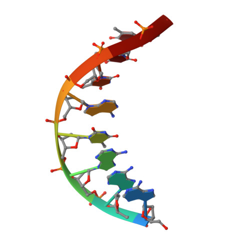
Structure of a DNA duplex with all-AT base pairs.

Valls, N.Richter, M.Subirana, J.A.

(2005) Acta Crystallogr D Biol Crystallogr 61: 1587-1593

  • DOI: https://doi.org/10.1107/S0907444905029781
  • Primary Citation Related Structures: 
    2A2T

  • PubMed Abstract: 

    The structure of the oligonucleotide duplex d(AAATATTT)(2) has been obtained in two crystal forms. In both cases the duplexes show Watson-Crick base pairs and are organized as a helical arrangement of stacked oligonucleotides. The helices contain either 11 duplexes in seven turns (form I) or 14 duplexes in nine turns (form II). As a result, the crystals have rather large unit cells. Such an organization of DNA duplexes has not been previously described in oligonucleotide crystal structures. The columns of stacked duplexes are organized in a pseudohexagonal arrangement. They do not show any direct lateral interactions; instead, these are indirectly mediated by shared and disordered hydrated ions. Such interactions are similar to those found in DNA fibres. The structural parameters of the individual base steps coincide with those found in other structures which contain CG terminal base pairs.


  • Organizational Affiliation
    • Departament d'Enginyeria Química, ETSEIB, Universitat Politècnica de Catalunya, Barclelona, Spain.

Macromolecule Content 

  • Total Structure Weight: 27.82 kDa 
  • Atom Count: 1,823 
  • Modeled Residue Count: 88 
  • Deposited Residue Count: 88 
  • Unique nucleic acid chains: 1

Macromolecules

Find similar nucleic acids by:  Sequence
Entity ID: 1
MoleculeChains LengthOrganismImage
5'-D(*AP*AP*AP*TP*AP*TP*TP*T)-3'8N/A
Sequence Annotations
Expand
Reference Sequence

Small Molecules

Ligands 2 Unique
IDChains Name / Formula / InChI Key2D Diagram3D Interactions
CAC

Query on CAC


Download:Ideal Coordinates CCD File
R [auth E]
S [auth E]
T [auth E]
U [auth E]
V [auth E]
R [auth E],
S [auth E],
T [auth E],
U [auth E],
V [auth E],
W [auth E]
CACODYLATE ION
C2 H6 As O2
OGGXGZAMXPVRFZ-UHFFFAOYSA-M
MN

Query on MN


Download:Ideal Coordinates CCD File
L [auth D]
M [auth E]
N [auth E]
O [auth E]
P [auth E]
L [auth D],
M [auth E],
N [auth E],
O [auth E],
P [auth E],
Q [auth E]
MANGANESE (II) ION
Mn
WAEMQWOKJMHJLA-UHFFFAOYSA-N

Experimental Data & Validation

Experimental Data

  • Method: X-RAY DIFFRACTION
  • Resolution: 3.10 Å
  • R-Value Free:  0.357 (Depositor), 0.339 (DCC) 
  • R-Value Work:  0.272 (Depositor) 
  • R-Value Observed: 0.276 (Depositor) 
Space Group: C 1 2 1
Unit Cell:
Length ( Å )Angle ( ˚ )
a = 147.623α = 90
b = 24.985β = 90.51
c = 82.139γ = 90
Software Package:
Software NamePurpose
REFMACrefinement
HKL-2000data reduction
SCALEPACKdata scaling
MOLREPphasing

Structure Validation

View Full Validation Report


Entry History 

Deposition Data

Revision History  (Full details and data files)

  • Version 1.0: 2005-11-29
    Type: Initial release
  • Version 1.1: 2008-04-30
    Changes: Version format compliance
  • Version 1.2: 2011-07-13
    Changes: Non-polymer description, Version format compliance
  • Version 1.3: 2024-03-13
    Changes: Data collection, Database references, Derived calculations