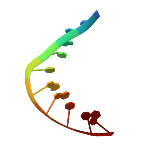
Solution structure of mithramycin dimers bound to partially overlapping sites on DNA.
Sastry, M., Fiala, R., Patel, D.J.(1995) J Mol Biol 251: 674-689
- PubMed: 7666419 Search on PubMed
- DOI: https://doi.org/10.1006/jmbi.1995.0464
- Primary Citation Related Structures:
207D - PubMed Abstract:
Mithramycin (MTH) is a DNA-binding antitumor agent containing A-B disaccharide and C-D-E trisaccharide segments projecting from opposite ends of an aglycone chromophore. We have previously reported on the solution structure of the MTH-DNA 6-mer complex based on a combined NMR and molecular dynamics study. This study established that the Mg(2+)-coordinated mithramycin dimer bound to a widened minor groove centered about the sequence-specific (G-C).(G-C) site and that the C-D-E trisaccharide segments from individual monomers were directed towards opposite ends of the helix spanning a six base-pair segment. This research is now extended to the binding of mithramycin dimers to partially overlapping sites on the self-complementary d(T-A-G-C-T-A-G-C-T-A) 10-mer duplex. The six base-pair mithramycin dimer footprint centered about (G-C).(G-C) steps should result in a potential steric clash in the center of the helix involving the inwardly pointing E-sugars of the pair of mithramycin dimers bound to the DNA 10-mer duplex. The MTH-d(T-A-G-C-T-A-G-C-T-A) complex (two MTH dimers per duplex) yields narrow and well-resolved NMR spectra, which have been assigned to identify intramolecular and intermolecular nuclear Overhauser enhancement (NOE) connectivities in the complex. The solution structure of the MTH-DNA 10-mer complex based on distance-restrained molecular dynamics calculations has defined the conformation of the drug and the DNA necessary for accommodation of the pair of mithramycin dimers on the DNA 10-mer helix. Specifically, the inwardly pointing E-sugars retain their face-down alignment towards the floor of the minor groove and occupy adjacent binding sites in the center of the duplex. This is achieved, in part, through torsion angle differences in the glycosidic linkage bonds along the length of the inwardly pointing aglycone-C-D-E trisaccharide segment relative to its outwardly pointing aglycone-C-D-E trisaccharide counterpart in the complex. In addition, a pronounced kink at the central (T-A).(T-A) step opens the minor groove and generates additional space to accommodate the inwardly pointing E-sugars at adjacent sites in the MTH-DNA 10-mer complex. These studies establish conformational plasticity in the C-D-E trisaccharide segment of the mithramycin dimer and deformability of the DNA helix allowing mithramycin dimers to bind to partially overlapping minor groove sites on the DNA helix.
- Department of Biochemistry and Molecular Biophysics, College of Physicians and Surgeons, Columbia University, New York, NY 10032, USA.
Organizational Affiliation: