1ZYE

Crystal structure analysis of Bovine Mitochondrial Peroxiredoxin III


Experimental Data Snapshot

  • Method: X-RAY DIFFRACTION
  • Resolution: 3.30 Å
  • R-Value Free: 0.265 
  • R-Value Work: 0.226 
  • R-Value Observed: 0.228 

wwPDB Validation   3D Report Full Report


This is version 1.5 of the entry. See complete history


Literature

Bovine Mitochondrial Peroxiredoxin III Forms a Two-Ring Catenane

Cao, Z.Roszak, A.W.Gourlay, L.J.Lindsay, J.G.Isaacs, N.W.

(2005) Structure 13: 1661-1664

  • DOI: https://doi.org/10.1016/j.str.2005.07.021
  • Primary Citation of Related Structures:  
    1ZYE

  • PubMed Abstract: 

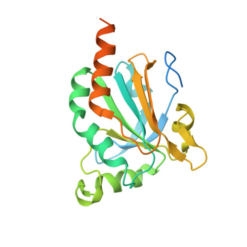
    A crystal structure is reported for the C168S mutant of a typical 2-Cys peroxiredoxin III (Prx III) from bovine mitochondria at a resolution of 3.3 A. Prx III is present as a two-ring catenane comprising two interlocking dodecameric toroids that are assembled from basic dimeric units. Each ring has an external diameter of 150 A and encompasses a central cavity that is 70 A in width. The concatenated dodecamers are inclined at an angle of 55 degrees, which provides a large contact surface between the rings. Dimer-dimer contacts involved in toroid formation are hydrophobic in nature, whereas the 12 areas of contact between interlocked rings arise from polar interactions. These two major modes of subunit interaction provide important insights into possible mechanisms of catenane formation.


  • Organizational Affiliation

    Department of Biochemistry and Molecular Biology, University of Glasgow, Glasgow G12 8QQ, United Kingdom.


Macromolecules
Find similar proteins by:  (by identity cutoff)  |  3D Structure
Entity ID: 1
MoleculeChains Sequence LengthOrganismDetailsImage
Thioredoxin-dependent peroxide reductase
A, B, C, D, E
A, B, C, D, E, F, G, H, I, J, K, L
220Bos taurusMutation(s): 1 
EC: 1.11.1.15
UniProt
Find proteins for P35705 (Bos taurus)
Explore P35705 
Go to UniProtKB:  P35705
Entity Groups  
Sequence Clusters30% Identity50% Identity70% Identity90% Identity95% Identity100% Identity
UniProt GroupP35705
Sequence Annotations
Expand
  • Reference Sequence
Experimental Data & Validation

Experimental Data

  • Method: X-RAY DIFFRACTION
  • Resolution: 3.30 Å
  • R-Value Free: 0.265 
  • R-Value Work: 0.226 
  • R-Value Observed: 0.228 
  • Space Group: C 1 2 1
Unit Cell:
Length ( Å )Angle ( ˚ )
a = 300.995α = 90
b = 80.679β = 112.76
c = 124.295γ = 90
Software Package:
Software NamePurpose
REFMACrefinement
MOSFLMdata reduction
CCP4data scaling
PHASERphasing

Structure Validation

View Full Validation Report


Entry History 

Deposition Data

Revision History  (Full details and data files)

  • Version 1.0: 2005-09-20
    Type: Initial release
  • Version 1.1: 2008-04-30
    Changes: Version format compliance
  • Version 1.2: 2011-07-13
    Changes: Derived calculations, Version format compliance
  • Version 1.3: 2020-09-09
    Changes: Database references, Structure summary
  • Version 1.4: 2021-11-10
    Changes: Database references
  • Version 1.5: 2023-10-25
    Changes: Data collection, Refinement description