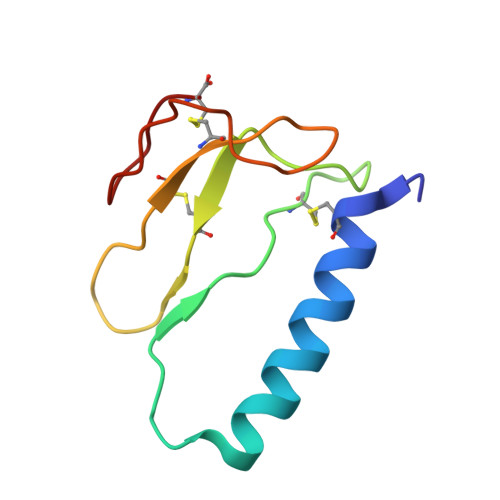
1ZT3 | pdb_00001zt3

C-terminal domain of Insulin-like Growth Factor Binding Protein-1 isolated from human amniotic fluid


Experimental Data Snapshot

  • Method: X-RAY DIFFRACTION
  • Resolution: 1.80 Å
  • R-Value Free: 
    0.275 (Depositor), 0.269 (DCC) 
  • R-Value Work: 
    0.221 (Depositor), 0.218 (DCC) 
  • R-Value Observed: 
    0.224 (Depositor) 

wwPDB Validation 3D Report Full Report

Validation slider image for 1ZT3

This is version 1.4 of the entry. See complete history. 

Literature

Structure and Properties of the C-terminal Domain of Insulin-like Growth Factor-binding Protein-1 Isolated from Human Amniotic Fluid

Sala, A., Capaldi, S., Campagnoli, M., Faggion, B., Labo, S., Perduca, M., Romano, A., Carrizo, M.E., Valli, M., Visai, L., Minchiotti, L., Galliano, M., Monaco, H.L.

(2005) J Biol Chem 280: 29812-29819

  • DOI: https://doi.org/10.1074/jbc.M504304200
  • Primary Citation Related Structures: 
    1ZT3, 1ZT5

  • PubMed Abstract: 

    Insulin-like growth factor (IGF)-binding protein-1 (IGFBP-1) regulates the activity of the insulin-like growth factors in early pregnancy and is, thus, thought to play a key role at the fetal-maternal interface. The C-terminal domain of IGFBP-1 and three isoforms of the intact protein were isolated from human amniotic fluid, and sequencing of the four N-terminal polypeptide chains showed them to be highly pure. The addition of both intact IGFBP-1 and its C-terminal fragment to cultured fibroblasts has a similar stimulating effect on cell migration, and therefore, the domain has a biological activity on its own. The three-dimensional structure of the C-terminal domain was determined by x-ray crystallography to 1.8 Angstroms resolution. The fragment folds as a thyroglobulin type I domain and was found to bind the Fe(2+) ion in the crystals through the only histidine residue present in the polypeptide chain. Iron (II) decreases the binding of intact IGFBP-1 and the C-terminal domain to IGF-II, suggesting that the metal binding site is close to or part of the surface of interaction of the two molecules.


  • Organizational Affiliation: 
    • Department of Biochemistry A. Castellani, University of Pavia, Italy.

Macromolecule Content 

  • Total Structure Weight: 9.27 kDa 
  • Atom Count: 690 
  • Modeled Residue Count: 80 
  • Deposited Residue Count: 80 
  • Unique protein chains: 1

Macromolecules

Find similar proteins by:|  3D Structure
Entity ID: 1
MoleculeChains  Sequence LengthOrganismDetailsImage
Insulin-like growth factor binding protein 180Homo sapiensMutation(s): 0 
UniProt & NIH Common Fund Data Resources
Find proteins for P08833 (Homo sapiens)
Explore P08833 
Go to UniProtKB:  P08833
PHAROS:  P08833
GTEx:  ENSG00000146678 
Entity Groups
Sequence Clusters30% Identity50% Identity70% Identity90% Identity95% Identity100% Identity
UniProt GroupP08833
Sequence Annotations
Expand
Reference Sequence

Small Molecules

Ligands 1 Unique
IDChains Name / Formula / InChI Key2D Diagram3D Interactions
DIO

Query on DIO


Download:Ideal Coordinates CCD File
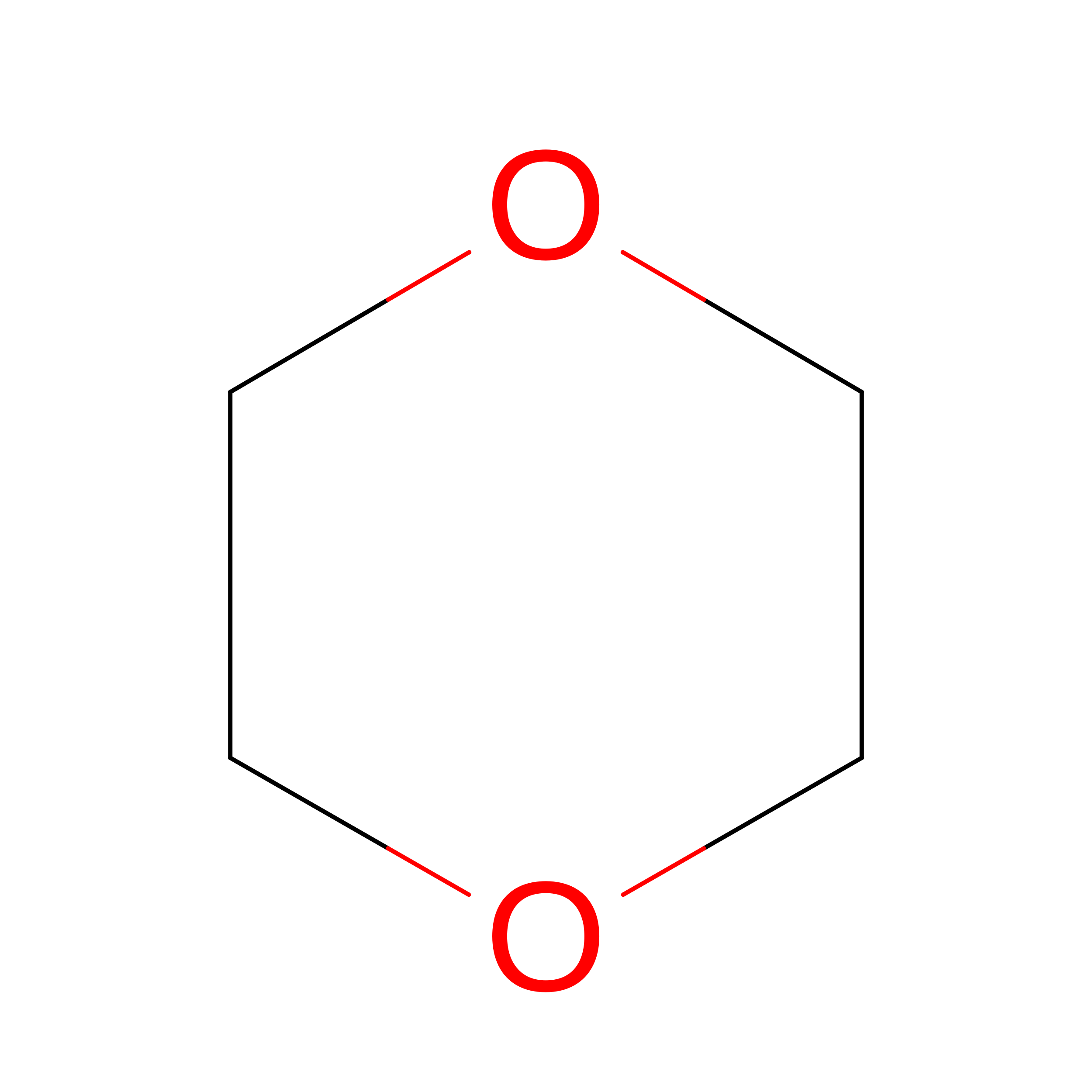
B [auth A]1,4-DIETHYLENE DIOXIDE
C4 H8 O2
RYHBNJHYFVUHQT-UHFFFAOYSA-N

Experimental Data & Validation

Experimental Data

  • Method: X-RAY DIFFRACTION
  • Resolution: 1.80 Å
  • R-Value Free:  0.275 (Depositor), 0.269 (DCC) 
  • R-Value Work:  0.221 (Depositor), 0.218 (DCC) 
  • R-Value Observed: 0.224 (Depositor) 
Space Group: P 21 21 2
Unit Cell:
Length ( Å )Angle ( ˚ )
a = 38.54α = 90
b = 60.39β = 90
c = 31.24γ = 90
Software Package:
Software NamePurpose
REFMACrefinement
MAR345data collection
AUTOMARdata reduction
SHARPphasing

Structure Validation

View Full Validation Report


Entry History 

Revision History  (Full details and data files)

  • Version 1.0: 2005-06-28
    Type: Initial release
  • Version 1.1: 2008-04-30
    Changes: Version format compliance
  • Version 1.2: 2011-07-13
    Changes: Version format compliance
  • Version 1.3: 2017-10-11
    Changes: Refinement description
  • Version 1.4: 2024-10-16
    Changes: Data collection, Database references, Derived calculations, Structure summary