1ZM9

Structure of eEF2-ETA in complex with PJ34


Experimental Data Snapshot

  • Method: X-RAY DIFFRACTION
  • Resolution: 2.80 Å
  • R-Value Free: 0.274 
  • R-Value Work: 0.254 
  • R-Value Observed: 0.255 

wwPDB Validation   3D Report Full Report


Ligand Structure Quality Assessment 


This is version 1.2 of the entry. See complete history


Literature

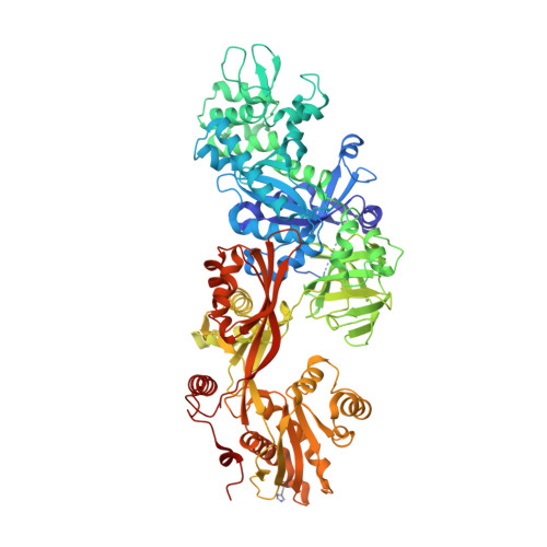
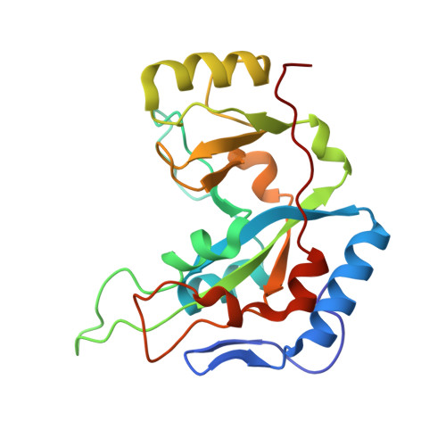
Exotoxin A-eEF2 complex structure indicates ADP ribosylation by ribosome mimicry.

Joergensen, R.Merrill, A.R.Yates, S.P.Marquez, V.E.Schwan, A.L.Boesen, T.Andersen, G.R.

(2005) Nature 436: 979-984

  • DOI: https://doi.org/10.1038/nature03871
  • Primary Citation of Related Structures:  
    1ZM2, 1ZM3, 1ZM4, 1ZM9

  • PubMed Abstract: 
  • The bacteria causing diphtheria, whooping cough, cholera and other diseases secrete mono-ADP-ribosylating toxins that modify intracellular proteins. Here, we describe four structures of a catalytically active complex between a fragment of Pseudomonas aeruginosa exotoxin A (ETA) and its protein substrate, translation elongation factor 2 (eEF2) ...

    The bacteria causing diphtheria, whooping cough, cholera and other diseases secrete mono-ADP-ribosylating toxins that modify intracellular proteins. Here, we describe four structures of a catalytically active complex between a fragment of Pseudomonas aeruginosa exotoxin A (ETA) and its protein substrate, translation elongation factor 2 (eEF2). The target residue in eEF2, diphthamide (a modified histidine), spans across a cleft and faces the two phosphates and a ribose of the non-hydrolysable NAD+ analogue, betaTAD. This suggests that the diphthamide is involved in triggering NAD+ cleavage and interacting with the proposed oxacarbenium intermediate during the nucleophilic substitution reaction, explaining the requirement of diphthamide for ADP ribosylation. Diphtheria toxin may recognize eEF2 in a manner similar to ETA. Notably, the toxin-bound betaTAD phosphates mimic the phosphate backbone of two nucleotides in a conformational switch of 18S rRNA, thereby achieving universal recognition of eEF2 by ETA.


    Organizational Affiliation

    Centre for Structural Biology, Department of Molecular Biology, University of Aarhus, Gustav Wieds Vej 10C, DK-8000, Denmark.


Macromolecules
Find similar proteins by:  (by identity cutoff)  |  3D Structure
Entity ID: 1
MoleculeChainsSequence LengthOrganismDetailsImage
Elongation factor 2A, C, E842Saccharomyces cerevisiaeMutation(s): 1 
UniProt
Find proteins for P32324 (Saccharomyces cerevisiae (strain ATCC 204508 / S288c))
Explore P32324 
Go to UniProtKB:  P32324
Entity Groups  
Sequence Clusters30% Identity50% Identity70% Identity90% Identity95% Identity100% Identity
UniProt GroupP32324
Protein Feature View
Expand
  • Reference Sequence
Find similar proteins by:  (by identity cutoff)  |  3D Structure
Entity ID: 2
MoleculeChainsSequence LengthOrganismDetailsImage
exotoxin AB, D, F207Pseudomonas aeruginosaMutation(s): 0 
EC: 2.4.2.36
UniProt
Find proteins for P11439 (Pseudomonas aeruginosa (strain ATCC 15692 / DSM 22644 / CIP 104116 / JCM 14847 / LMG 12228 / 1C / PRS 101 / PAO1))
Explore P11439 
Go to UniProtKB:  P11439
Entity Groups  
Sequence Clusters30% Identity50% Identity70% Identity90% Identity95% Identity100% Identity
UniProt GroupP11439
Protein Feature View
Expand
  • Reference Sequence
Small Molecules
Ligands 1 Unique
IDChainsName / Formula / InChI Key2D Diagram3D Interactions
P34
Query on P34

Download Ideal Coordinates CCD File 
G [auth B],
H [auth D],
I [auth F]
N~2~,N~2~-DIMETHYL-N~1~-(6-OXO-5,6-DIHYDROPHENANTHRIDIN-2-YL)GLYCINAMIDE
C17 H17 N3 O2
UYJZZVDLGDDTCL-UHFFFAOYSA-N
 Ligand Interaction
Modified Residues  1 Unique
IDChainsTypeFormula2D DiagramParent
DDE
Query on DDE
A, C, E L-PEPTIDE LINKINGC13 H24 N5 O3HIS
Experimental Data & Validation

Experimental Data

  • Method: X-RAY DIFFRACTION
  • Resolution: 2.80 Å
  • R-Value Free: 0.274 
  • R-Value Work: 0.254 
  • R-Value Observed: 0.255 
  • Space Group: C 1 2 1
Unit Cell:
Length ( Å )Angle ( ˚ )
a = 330.84α = 90
b = 68.74β = 103.5
c = 191.46γ = 90
Software Package:
Software NamePurpose
XDSdata scaling
XDSdata reduction
MOLREPphasing
CNSrefinement

Structure Validation

View Full Validation Report


Ligand Structure Quality Assessment 


Entry History 

Deposition Data

Revision History  (Full details and data files)

  • Version 1.0: 2005-05-24
    Type: Initial release
  • Version 1.1: 2008-04-30
    Changes: Version format compliance
  • Version 1.2: 2011-07-13
    Changes: Version format compliance