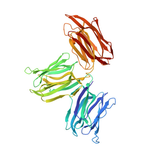
1ZGR

Crystal structure of the Parkia platycephala seed lectin


Experimental Data Snapshot

  • Method: X-RAY DIFFRACTION
  • Resolution: 2.50 Å
  • R-Value Free: 0.243 
  • R-Value Work: 0.214 
  • R-Value Observed: 0.215 

wwPDB Validation   3D Report Full Report


This is version 1.4 of the entry. See complete history


Literature

The first crystal structure of a mimosoideae lectin reveals a novel quaternary arrangement of a widespread domain.

Gallego Del Sol, F.Nagano, C.Cavada, B.S.Calvete, J.J.

(2005) J Mol Biol 353: 574-583

  • DOI: https://doi.org/10.1016/j.jmb.2005.08.055
  • Primary Citation of Related Structures:  
    1ZGR, 1ZGS

  • PubMed Abstract: 

    The crystal structures of the apo and mannose-bound Parkia platycephala seed lectin represent the first structure of a Mimosoideae lectin and a novel circular arrangement of beta-prism domains, and highlight the adaptability of the beta-prism fold as a building block in the evolution of plant lectins. The P.platycephala lectin is a dimer both in solution and in the crystals. Mannose binding to each of the three homologous carbohydrate-recognition domains of the lectin occurs through different modes, and restrains the flexibility of surface-exposed loops and residues involved in carbohydrate recognition. The planar array of carbohydrate-binding sites on the rim of the toroid-shaped structure of the P.platycephala lectin dimer immediately suggests a mechanism to promote multivalent interactions leading to cross-linking of carbohydrate ligands as part of the host strategy against phytopredators and pathogens. The cyclic structure of the P.platycephala lectin points to the convergent evolution of a structural principle for the construction of lectins involved in host defense or in attacking other organisms.


  • Organizational Affiliation

    Instituto de Biomedicina de Valencia, CSIC, E-46010 Valencia, Spain.


Macromolecules
Find similar proteins by:  (by identity cutoff)  |  3D Structure
Entity ID: 1
MoleculeChains Sequence LengthOrganismDetailsImage
Mannose/glucose-specific lectin
A, B
447Parkia platycephalaMutation(s): 0 
UniProt
Find proteins for P83304 (Parkia platycephala)
Explore P83304 
Go to UniProtKB:  P83304
Entity Groups  
Sequence Clusters30% Identity50% Identity70% Identity90% Identity95% Identity100% Identity
UniProt GroupP83304
Sequence Annotations
Expand
  • Reference Sequence
Experimental Data & Validation

Experimental Data

  • Method: X-RAY DIFFRACTION
  • Resolution: 2.50 Å
  • R-Value Free: 0.243 
  • R-Value Work: 0.214 
  • R-Value Observed: 0.215 
  • Space Group: P 21 21 21
Unit Cell:
Length ( Å )Angle ( ˚ )
a = 63.597α = 90
b = 68.511β = 90
c = 208.531γ = 90
Software Package:
Software NamePurpose
REFMACrefinement
MOSFLMdata reduction
CCP4data scaling
AMoREphasing

Structure Validation

View Full Validation Report


Entry History 

Deposition Data

Revision History  (Full details and data files)

  • Version 1.0: 2005-10-11
    Type: Initial release
  • Version 1.1: 2008-04-30
    Changes: Version format compliance
  • Version 1.2: 2011-07-13
    Changes: Version format compliance
  • Version 1.3: 2019-11-06
    Changes: Advisory, Data collection
  • Version 1.4: 2024-03-13
    Changes: Advisory, Data collection, Database references