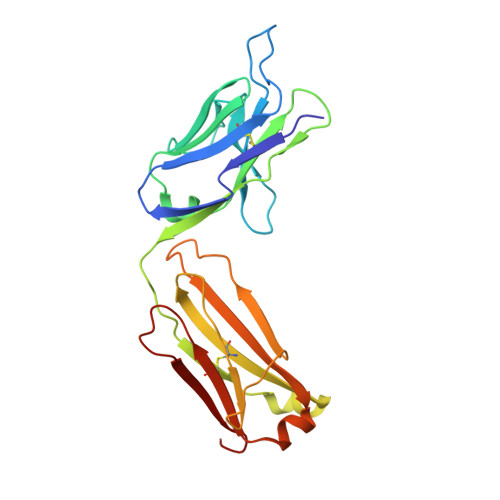
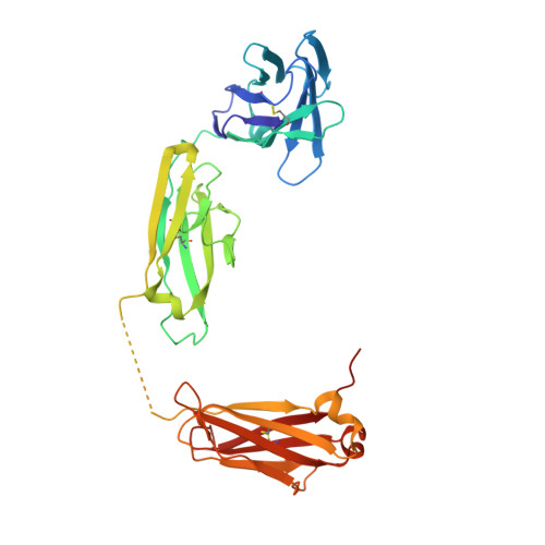
The Structure of an Antitumor C(H)2-domain-deleted Humanized Antibody.
Larson, S.B., Day, J.S., Glaser, S., Braslawsky, G., McPherson, A.(2005) J Mol Biology 348: 1177-1190
- PubMed: 15854653 Search on PubMed
- DOI: https://doi.org/10.1016/j.jmb.2005.03.036
- Primary Citation Related Structures:
1ZA6 - PubMed Abstract:
C(H)2-domain-deleted CC49 (HuCC49DeltaCH2), a recombinant humanized antibody that recognizes the TAG-72 antigen expressed on a variety of human carcinomas, is secreted from cultured cells as a mixture of two homodimeric isoforms. Isoform A contains two covalent interchain disulfide bonds at heavy chain positions 239 and 242, while isoform B fails to develop any interchain disulfide bonds but has 239-242 intrachain disulfide bonds instead. Form A is currently in preclinical development as a therapeutic agent for treating colorectal carcinoma, though form B shows equal efficacy. HuCC49DeltaCH2 form B can be crystallized from sodium formate only in the presence of detergents. X-ray diffraction data were collected on a single cryo-cooled crystal grown with Triton X-100 and the structure was solved by molecular replacement. The model has refined to R=0.246 (R(free)=0.297) for 2.8A data. The antibodies pack in the crystal around crystallographic 2-fold axes as tetramers with approximate 222 symmetry. Atomic force microscopy studies show that this tetrameric structure is the crystal building block and also exists free in the mother liquor. The tetramer is composed of two rings, back-to-back, with a thickness of approximately 83A. Each ring is composed of two antibodies with the complementarity-determining regions (CDR) of the two Fabs of one antibody interacting with the CDR regions of the second antibody in a head-to-head fashion. These rings are approximately 167A long and 112A wide. The C(H)3 domain is inverted with respect to the Fabs when compared to the usual orientation found in conventional antibodies. The polypeptides joining the C(H)3 domains to the Fab portions of the antibody are not seen and are almost certainly disordered. The antigen combining site of HuCC49DeltaCH2 is very similar, but not identical, in topology and charge distribution to that of antibody B72.3, which binds a similar epitope on TAG-72. The combining site consists of a deep cleft, heavily lined with aromatic amino acid side-chains but bounded by numerous charged groups.
- Department of Molecular Biology and Biochemistry, The University of California, Irvine, CA 92697-3900, USA.
Organizational Affiliation: