1ZA1

Structure of wild-type E. coli Aspartate Transcarbamoylase in the presence of CTP at 2.20 A resolution


Experimental Data Snapshot

  • Method: X-RAY DIFFRACTION
  • Resolution: 2.20 Å
  • R-Value Free: 0.252 
  • R-Value Work: 0.205 
  • R-Value Observed: 0.205 

wwPDB Validation   3D Report Full Report


Ligand Structure Quality Assessment 


This is version 1.4 of the entry. See complete history


Literature

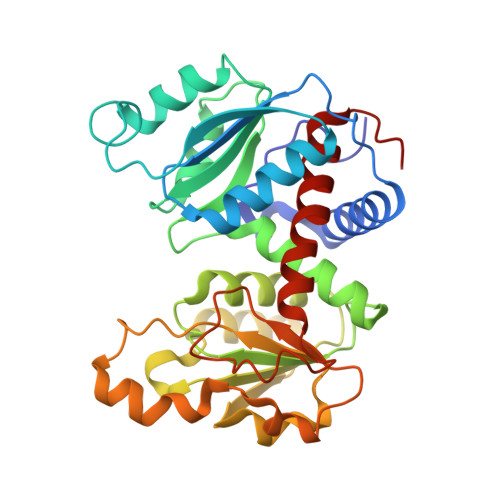
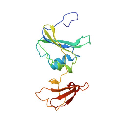
Structural basis for ordered substrate binding and cooperativity in aspartate transcarbamoylase

Wang, J.Stieglitz, K.A.Cardia, J.P.Kantrowitz, E.R.

(2005) Proc Natl Acad Sci U S A 102: 8881-8886

  • DOI: https://doi.org/10.1073/pnas.0503742102
  • Primary Citation of Related Structures:  
    1ZA1, 1ZA2

  • PubMed Abstract: 

    X-ray structures of aspartate transcarbamoylase in the absence and presence of the first substrate carbamoyl phosphate are reported. These two structures in conjunction with in silico docking experiments provide snapshots of critical events in the function of the enzyme. The ordered substrate binding, observed experimentally, can now be structurally explained by a conformational change induced upon the binding of carbamoyl phosphate. This induced fit dramatically alters the electrostatics of the active site, creating a binding pocket for aspartate. Upon aspartate binding, a further change in electrostatics causes a second induced fit, the domain closure. This domain closure acts as a clamp that both facilitates catalysis by approximation and also initiates the global conformational change that manifests homotropic cooperativity.


  • Organizational Affiliation

    Department of Chemistry, Boston College, Merkert Chemistry Center, Chestnut Hill, MA 02467, USA.


Macromolecules
Find similar proteins by:  (by identity cutoff)  |  3D Structure
Entity ID: 1
MoleculeChains Sequence LengthOrganismDetailsImage
Aspartate carbamoyltransferase catalytic chain
A, C
310Escherichia coliMutation(s): 0 
Gene Names: PYRB
EC: 2.1.3.2
UniProt
Find proteins for P0A786 (Escherichia coli (strain K12))
Explore P0A786 
Go to UniProtKB:  P0A786
Entity Groups  
Sequence Clusters30% Identity50% Identity70% Identity90% Identity95% Identity100% Identity
UniProt GroupP0A786
Sequence Annotations
Expand
  • Reference Sequence
Find similar proteins by:  (by identity cutoff)  |  3D Structure
Entity ID: 2
MoleculeChains Sequence LengthOrganismDetailsImage
Aspartate carbamoyltransferase regulatory chain
B, D
153Escherichia coliMutation(s): 0 
Gene Names: PYRI
EC: 2.1.3.2
UniProt
Find proteins for P0A7F3 (Escherichia coli (strain K12))
Explore P0A7F3 
Go to UniProtKB:  P0A7F3
Entity Groups  
Sequence Clusters30% Identity50% Identity70% Identity90% Identity95% Identity100% Identity
UniProt GroupP0A7F3
Sequence Annotations
Expand
  • Reference Sequence
Experimental Data & Validation

Experimental Data

  • Method: X-RAY DIFFRACTION
  • Resolution: 2.20 Å
  • R-Value Free: 0.252 
  • R-Value Work: 0.205 
  • R-Value Observed: 0.205 
  • Space Group: P 3 2 1
Unit Cell:
Length ( Å )Angle ( ˚ )
a = 120.294α = 90
b = 120.294β = 90
c = 142.555γ = 120
Software Package:
Software NamePurpose
CNSrefinement
CNSphasing

Structure Validation

View Full Validation Report


Ligand Structure Quality Assessment 


Entry History 

Deposition Data

Revision History  (Full details and data files)

  • Version 1.0: 2005-06-07
    Type: Initial release
  • Version 1.1: 2008-04-30
    Changes: Version format compliance
  • Version 1.2: 2011-07-13
    Changes: Derived calculations, Version format compliance
  • Version 1.3: 2017-10-11
    Changes: Refinement description
  • Version 1.4: 2023-08-23
    Changes: Data collection, Database references, Derived calculations, Refinement description