1YZP | pdb_00001yzp

Substrate-free manganese peroxidase


Experimental Data Snapshot

  • Method: X-RAY DIFFRACTION
  • Resolution: 1.60 Å
  • R-Value Free: 
    0.203 (Depositor) 
  • R-Value Work: 
    0.147 (Depositor) 
  • R-Value Observed: 
    0.153 (Depositor) 

Starting Model: experimental
View more details

wwPDB Validation 3D Report Full Report

Validation slider image for 1YZP

This is version 2.2 of the entry. See complete history

Literature

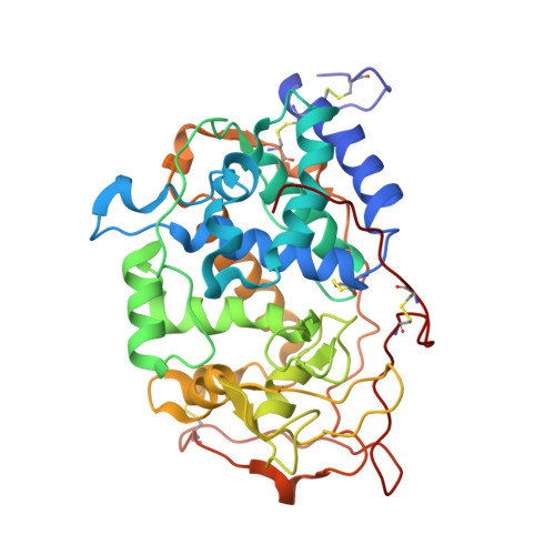
High-Resolution Crystal Structure of Manganese Peroxidase: Substrate and Inhibitor Complexes.

Sundaramoorthy, M.Youngs, H.L.Gold, M.H.Poulos, T.L.

(2005) Biochemistry 44: 6463-6470

  • DOI: https://doi.org/10.1021/bi047318e
  • Primary Citation Related Structures: 
    1YYD, 1YYG, 1YZP, 1YZR

  • PubMed Abstract: 

    Manganese peroxidase (MnP) is an extracellular heme enzyme that catalyzes the peroxide-dependent oxidation of Mn(II) to Mn(III). The Mn(III) is released from the enzyme in complex with oxalate. One heme propionate and the side chains of Glu35, Glu39, and Asp179 were identified as Mn(II) ligands in the 2.0 A resolution crystal structure. The new 1.45 A crystal structure of MnP complexed with Mn(II) provides a more accurate view of the Mn-binding site. New features include possible partial protonation of Glu39 in the Mn-binding site and glycosylation at Ser336. This is also the first report of MnP-inhibitor complex structures. At the Mn-binding site, divalent Cd(II) exhibits octahedral, hexacoordinate ligation geometry similar to that of Mn(II). Cd(II) also binds to a putative second weak metal-binding site with tetrahedral geometry at the C-terminus of the protein. Unlike that for Mn(II) and Cd(II), coordination of trivalent Sm(III) at the Mn-binding site is octacoordinate. Sm(III) was removed from a MnP-Sm(III) crystal by soaking the crystal in oxalate and then reintroduced into the binding site. Thus, direct comparisons of Sm(III)-bound and metal-free structures were made using the same crystal. No ternary complex was observed upon incubation with oxalate. The reversible binding of Sm(III) may be a useful model for the reversible binding of Mn(III) to the enzyme, which is too unstable to allow similar examination.


  • Organizational Affiliation
    • Division of Nephrology, Department of Medicine, Center for Matrix Biology, Vanderbilt University Medical Center, Nashville, Tennessee 37232-2372, USA. m.sundaramoorthy@ vanderbilt.edu

Macromolecule Content 

  • Total Structure Weight: 38.88 kDa 
  • Atom Count: 3,281 
  • Modeled Residue Count: 357 
  • Deposited Residue Count: 357 
  • Unique protein chains: 1

Macromolecules

Find similar proteins by:|  3D Structure
Entity ID: 1
MoleculeChains  Sequence LengthOrganismDetailsImage
Peroxidase manganese-dependent I357Phanerodontia chrysosporiumMutation(s): 0 
EC: 1.11.1.13
UniProt
Find proteins for Q02567 (Phanerodontia chrysosporium)
Explore Q02567 
Go to UniProtKB:  Q02567
Entity Groups
Sequence Clusters30% Identity50% Identity70% Identity90% Identity95% Identity100% Identity
UniProt GroupQ02567
Glycosylation
Glycosylation Sites: 2
Sequence Annotations
Expand
Reference Sequence

Oligosaccharides

Help  
Entity ID: 2
MoleculeChains Length2D Diagram GlycosylationD Interactions
2-acetamido-2-deoxy-beta-D-glucopyranose-(1-4)-2-acetamido-2-deoxy-beta-D-glucopyranose
B
2N-Glycosylation
Glycosylation Resources
GlyTouCan: G42666HT
GlyCosmos: G42666HT
GlyGen: G42666HT

Experimental Data & Validation

Experimental Data

  • Method: X-RAY DIFFRACTION
  • Resolution: 1.60 Å
  • R-Value Free:  0.203 (Depositor) 
  • R-Value Work:  0.147 (Depositor) 
  • R-Value Observed: 0.153 (Depositor) 
Space Group: C 1 2 1
Unit Cell:
Length ( Å )Angle ( ˚ )
a = 160.595α = 90
b = 45.356β = 97.25
c = 52.817γ = 90
Software Package:
Software NamePurpose
DENZOdata reduction
SCALEPACKdata scaling
SHELXLrefinement
SHELXL-97refinement

Structure Validation

View Full Validation Report


Entry History 

Deposition Data

Revision History  (Full details and data files)

  • Version 1.0: 2005-05-10
    Type: Initial release
  • Version 1.1: 2008-04-30
    Changes: Version format compliance
  • Version 1.2: 2011-07-13
    Changes: Non-polymer description, Version format compliance
  • Version 1.3: 2018-01-24
    Changes: Database references
  • Version 2.0: 2020-07-29
    Type: Remediation
    Reason: Carbohydrate remediation
    Changes: Atomic model, Data collection, Derived calculations, Structure summary
  • Version 2.1: 2023-08-23
    Changes: Data collection, Database references, Refinement description, Structure summary
  • Version 2.2: 2024-11-20
    Changes: Structure summary