1YYF

Correction of X-ray Intensities from an HslV-HslU co-crystal containing lattice translocation defects


Experimental Data Snapshot

  • Method: X-RAY DIFFRACTION
  • Resolution: 4.16 Å
  • R-Value Free: 0.346 
  • R-Value Work: 0.277 
  • R-Value Observed: 0.280 

wwPDB Validation   3D Report Full Report


Ligand Structure Quality Assessment 


This is version 1.3 of the entry. See complete history


Literature

Correction of X-ray intensities from an HslV-HslU co-crystal containing lattice-translocation defects.

Wang, J.Rho, S.H.Park, H.H.Eom, S.H.

(2005) Acta Crystallogr D Biol Crystallogr 61: 932-941

  • DOI: https://doi.org/10.1107/S0907444905009546
  • Primary Citation of Related Structures:  
    1YYF

  • PubMed Abstract: 

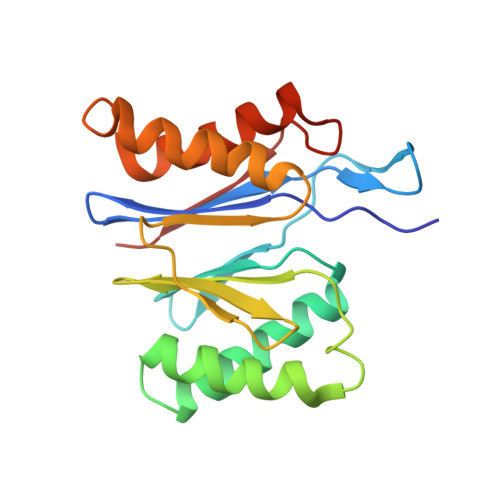
    Because of lattice-translocation defects, two identical but translated lattices can coexist as a single coherent mosaic block in a crystal. The observed structure in such cases is a weighted sum of two identical but translated structures, one from each lattice; the observed structure factors are a weighted vector sum of the structure factors with identical unit amplitudes but shifted phases. The correction of X-ray intensities from a single crystal containing these defects of the hybrid HslV-HslU complex, which consists of Escherichia coli HslU and Bacillus subtilis HslV (also known as CodW), is reported. When intensities are not corrected, a biologically irrelevant complex (with CodW from one lattice and HslU from another) is implied to exist. Only upon correction does a biologically functional CodW-HslU complex structure emerge.


  • Organizational Affiliation

    Department of Molecular Biophysics and Biochemistry, Yale University, 266 Whitney Avenue, New Haven, CT 06520-8114, USA. wang@mail.csb.yale.edu


Macromolecules
Find similar proteins by:  (by identity cutoff)  |  3D Structure
Entity ID: 1
MoleculeChains Sequence LengthOrganismDetailsImage
ATP-dependent hsl protease ATP-binding subunit hslU
A, B
443Escherichia coliMutation(s): 0 
Gene Names: hslUhtpI
UniProt
Find proteins for P0A6H5 (Escherichia coli (strain K12))
Explore P0A6H5 
Go to UniProtKB:  P0A6H5
Entity Groups  
Sequence Clusters30% Identity50% Identity70% Identity90% Identity95% Identity100% Identity
UniProt GroupP0A6H5
Sequence Annotations
Expand
  • Reference Sequence
Find similar proteins by:  (by identity cutoff)  |  3D Structure
Entity ID: 2
MoleculeChains Sequence LengthOrganismDetailsImage
ATP-dependent protease hslVC [auth D],
D [auth C]
181Bacillus subtilisMutation(s): 0 
Gene Names: hslVclpQcodW
EC: 3.4.25
UniProt
Find proteins for P39070 (Bacillus subtilis (strain 168))
Explore P39070 
Go to UniProtKB:  P39070
Entity Groups  
Sequence Clusters30% Identity50% Identity70% Identity90% Identity95% Identity100% Identity
UniProt GroupP39070
Sequence Annotations
Expand
  • Reference Sequence
Small Molecules
Experimental Data & Validation

Experimental Data

  • Method: X-RAY DIFFRACTION
  • Resolution: 4.16 Å
  • R-Value Free: 0.346 
  • R-Value Work: 0.277 
  • R-Value Observed: 0.280 
  • Space Group: H 3 2
Unit Cell:
Length ( Å )Angle ( ˚ )
a = 181.196α = 90
b = 181.196β = 90
c = 529.924γ = 120
Software Package:
Software NamePurpose
REFMACrefinement
HKL-2000data reduction
SCALEPACKdata scaling
CNSphasing

Structure Validation

View Full Validation Report


Ligand Structure Quality Assessment 


Entry History 

Deposition Data

Revision History  (Full details and data files)

  • Version 1.0: 2005-07-12
    Type: Initial release
  • Version 1.1: 2008-04-30
    Changes: Version format compliance
  • Version 1.2: 2011-07-13
    Changes: Version format compliance
  • Version 1.3: 2024-02-14
    Changes: Data collection, Database references, Derived calculations, Refinement description