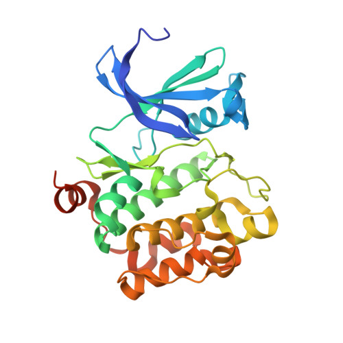
Crystal Structures of Proto-oncogene Kinase Pim1: A Target of Aberrant Somatic Hypermutations in Diffuse Large Cell Lymphoma.
Kumar, A., Mandiyan, V., Suzuki, Y., Zhang, C., Rice, J., Tsai, J., Artis, D.R., Ibrahim, P., Bremer, R.(2005) J Mol Biology 348: 183-193
- PubMed: 15808862
- DOI: https://doi.org/10.1016/j.jmb.2005.02.039
- Primary Citation of Related Structures:
1YWV, 1YXS, 1YXT, 1YXU, 1YXV, 1YXX - PubMed Abstract:
Pim1, a serine/threonine kinase, is involved in several biological functions including cell survival, proliferation, and differentiation. While pim1 has been shown to be involved in several hematopoietic cancers, it was also recently identified as a target of aberrant somatic hypermutation in diffuse large cell lymphoma (DLCL), the most common form of non-Hodgkin's lymphoma. The crystal structures of Pim1 in apo form and bound with AMPPNP have been solved and several unique features of Pim1 were identified, including the presence of an extra beta-hairpin in the N-terminal lobe and an unusual conformation of the hinge connecting the two lobes of the enzyme. While the apo Pim1 structure is nearly identical with that reported recently, the structure of AMPPNP bound to Pim1 is significantly different. Pim1 is unique among protein kinases due to the presence of a proline residue at position 123 that precludes the formation of the canonical second hydrogen bond between the hinge backbone and the adenine moiety of ATP. One crystal structure reported here shows that changing P123 to methionine, a common residue that offers the backbone hydrogen bond to ATP, does not restore the ATP binding pocket of Pim1 to that of a typical kinase. These unique structural features in Pim1 result in novel binding modes of AMP and a known kinase inhibitor scaffold, as shown by co-crystallography. In addition, the kinase activities of five Pim1 mutants identified in DLCL patients have been determined. In each case, the observed effects on kinase activity are consistent with the predicted consequences of the mutation on the Pim1 structure. Finally, 70 co-crystal structures of low molecular mass, low-affinity compounds with Pim1 have been solved in order to identify novel chemical classes as potential Pim1 inhibitors. Based on the structural information, opportunities for optimization of one specific example are discussed.
- Plexxikon, Inc., 91 Bolivar Drive, Berkeley, CA 94710, USA.
Organizational Affiliation:


