1YVG | pdb_00001yvg

Structural analysis of the catalytic domain of tetanus neurotoxin


Experimental Data Snapshot

  • Method: X-RAY DIFFRACTION
  • Resolution: 2.60 Å
  • R-Value Free: 
    0.276 (Depositor), 0.273 (DCC) 
  • R-Value Work: 
    0.218 (Depositor), 0.217 (DCC) 

Starting Model: experimental
View more details

wwPDB Validation 3D Report Full Report

Validation slider image for 1YVG

This is version 1.4 of the entry. See complete history

Literature

Structural analysis of the catalytic domain of tetanus neurotoxin.

Rao, K.N.Kumaran, D.Binz, T.Swaminathan, S.

(2005) Toxicon 45: 929-939

  • DOI: https://doi.org/10.1016/j.toxicon.2005.02.032
  • Primary Citation Related Structures: 
    1YVG

  • PubMed Abstract: 

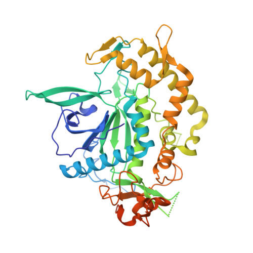
    Clostridium neurotoxins, comprising the tetanus neurotoxin and the seven antigenically distinct botulinum neurotoxins (BoNT/A-G), are among the known most potent bacterial protein toxins to humans. Although they have similar function, sequences and three-dimensional structures, the substrate specificity and the selectivity of peptide bond cleavage are different and unique. Tetanus and botulinum type B neurotoxins enzymatically cleave the same substrate, vesicle-associated membrane protein, at the same peptide bond though the optimum length of substrate peptide required for cleavage by them is different. Here, we present the first experimentally determined three-dimensional structure of the catalytic domain of tetanus neurotoxin and analyze its active site. The structure provides insight into the active site of tetanus toxin's proteolytic activity and the importance of the nucleophilic water and the role of the zinc ion. The probable reason for different modes of binding of vesicle-associated membrane protein to botulinum neurotoxin type B and the tetanus toxin is discussed. The structure provides a basis for designing a novel recombinant vaccine or structure-based drugs for tetanus.


  • Organizational Affiliation
    • Brookhaven National Laboratory, Biology Department, Upton, NY 11973, USA.

Macromolecule Content 

  • Total Structure Weight: 53.76 kDa 
  • Atom Count: 3,336 
  • Modeled Residue Count: 397 
  • Deposited Residue Count: 468 
  • Unique protein chains: 1

Macromolecules

Find similar proteins by:|  3D Structure
Entity ID: 1
MoleculeChains  Sequence LengthOrganismDetailsImage
Tetanus toxin, light chain468Clostridium tetaniMutation(s): 0 
Gene Names: tetX
EC: 3.4.24.68
UniProt
Find proteins for P04958 (Clostridium tetani (strain Massachusetts / E88))
Explore P04958 
Go to UniProtKB:  P04958
Entity Groups
Sequence Clusters30% Identity50% Identity70% Identity90% Identity95% Identity100% Identity
UniProt GroupP04958
Sequence Annotations
Expand
Reference Sequence

Small Molecules

Ligands 1 Unique
IDChains Name / Formula / InChI Key2D Diagram3D Interactions
ZN

Query on ZN


Download:Ideal Coordinates CCD File
B [auth A]ZINC ION
Zn
PTFCDOFLOPIGGS-UHFFFAOYSA-N

Experimental Data & Validation

Experimental Data

  • Method: X-RAY DIFFRACTION
  • Resolution: 2.60 Å
  • R-Value Free:  0.276 (Depositor), 0.273 (DCC) 
  • R-Value Work:  0.218 (Depositor), 0.217 (DCC) 
Space Group: C 2 2 2
Unit Cell:
Length ( Å )Angle ( ˚ )
a = 106.61α = 90
b = 177.28β = 90
c = 54.55γ = 90
Software Package:
Software NamePurpose
CBASSdata collection
SCALEPACKdata scaling
SOLVEphasing
CNSrefinement
SHARPphasing

Structure Validation

View Full Validation Report


Entry History 

Deposition Data

Revision History  (Full details and data files)

  • Version 1.0: 2005-03-22
    Type: Initial release
  • Version 1.1: 2008-04-30
    Changes: Version format compliance
  • Version 1.2: 2011-07-13
    Changes: Version format compliance
  • Version 1.3: 2011-11-16
    Changes: Atomic model
  • Version 1.4: 2023-08-23
    Changes: Data collection, Database references, Derived calculations, Refinement description