1YVB | pdb_00001yvb

the Plasmodium falciparum Cysteine Protease Falcipain-2


Experimental Data Snapshot

  • Method: X-RAY DIFFRACTION
  • Resolution: 2.70 Å
  • R-Value Free: 
    0.264 (Depositor), 0.264 (DCC) 
  • R-Value Work: 
    0.232 (Depositor), 0.231 (DCC) 
  • R-Value Observed: 
    0.232 (Depositor) 

Starting Model: experimental
View more details

wwPDB Validation 3D Report Full Report

Validation slider image for 1YVB

This is version 1.6 of the entry. See complete history

Literature

Structural basis for unique mechanisms of folding and hemoglobin binding by a malarial protease.

Wang, S.X.Pandey, K.C.Somoza, J.R.Sijwali, P.S.Kortemme, T.Brinen, L.S.Fletterick, R.J.Rosenthal, P.J.McKerrow, J.H.

(2006) Proc Natl Acad Sci U S A 103: 11503-11508

  • DOI: https://doi.org/10.1073/pnas.0600489103
  • Primary Citation Related Structures: 
    1YVB

  • PubMed Abstract: 

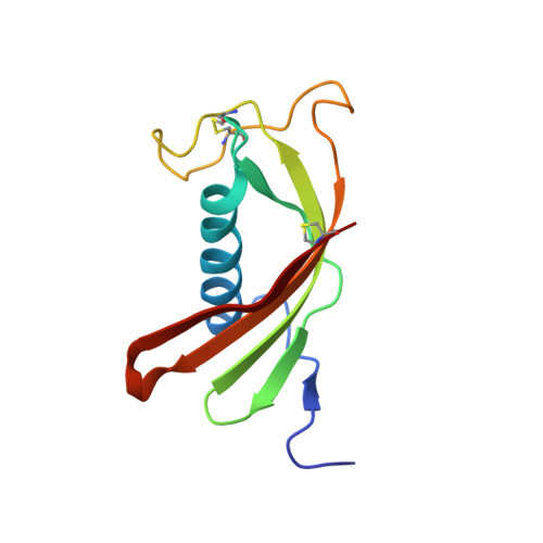
    Falcipain-2 (FP2), the major cysteine protease of the human malaria parasite Plasmodium falciparum, is a hemoglobinase and promising drug target. Here we report the crystal structure of FP2 in complex with a protease inhibitor, cystatin. The FP2 structure reveals two previously undescribed cysteine protease structural motifs, designated FP2(nose) and FP2(arm), in addition to details of the active site that will help focus inhibitor design. Unlike most cysteine proteases, FP2 does not require a prodomain but only the short FP2(nose) motif to correctly fold and gain catalytic activity. Our structure and mutagenesis data suggest a molecular basis for this unique mechanism by highlighting the functional role of two Tyr within FP2(nose) and a conserved Glu outside this motif. The FP2(arm) motif is required for hemoglobinase activity. The structure reveals topographic features and a negative charge cluster surrounding FP2(arm) that suggest it may serve as an exo-site for hemoglobin binding. Motifs similar to FP2(nose) and FP2(arm) are found only in related plasmodial proteases, suggesting that they confer malaria-specific functions.


  • Organizational Affiliation
    • Department of Pathology and the Sandler Center, San Francisco General Hospital, University of California, CA 94143, USA.

Macromolecule Content 

  • Total Structure Weight: 39.87 kDa 
  • Atom Count: 2,809 
  • Modeled Residue Count: 352 
  • Deposited Residue Count: 352 
  • Unique protein chains: 2

Macromolecules

Find similar proteins by:|  3D Structure
Entity ID: 1
MoleculeChains  Sequence LengthOrganismDetailsImage
falcipain 2241Plasmodium falciparum 3D7Mutation(s): 4 
Gene Names: AF239801
EC: 3.4.22
UniProt
Find proteins for Q8I6U4 (Plasmodium falciparum (isolate 3D7))
Explore Q8I6U4 
Go to UniProtKB:  Q8I6U4
Entity Groups
Sequence Clusters30% Identity50% Identity70% Identity90% Identity95% Identity100% Identity
UniProt GroupQ8I6U4
Sequence Annotations
Expand
Reference Sequence
Find similar proteins by:|  3D Structure
Entity ID: 2
MoleculeChains  Sequence LengthOrganismDetailsImage
CystatinB [auth I]111Gallus gallusMutation(s): 0 
UniProt
Find proteins for P01038 (Gallus gallus)
Explore P01038 
Go to UniProtKB:  P01038
Entity Groups
Sequence Clusters30% Identity50% Identity70% Identity90% Identity95% Identity100% Identity
UniProt GroupP01038
Sequence Annotations
Expand
Reference Sequence

Small Molecules

Ligands 1 Unique
IDChains Name / Formula / InChI Key2D Diagram3D Interactions
GOL

Query on GOL


Download:Ideal Coordinates CCD File
C [auth A]GLYCEROL
C3 H8 O3
PEDCQBHIVMGVHV-UHFFFAOYSA-N

Experimental Data & Validation

Experimental Data

  • Method: X-RAY DIFFRACTION
  • Resolution: 2.70 Å
  • R-Value Free:  0.264 (Depositor), 0.264 (DCC) 
  • R-Value Work:  0.232 (Depositor), 0.231 (DCC) 
  • R-Value Observed: 0.232 (Depositor) 
Space Group: P 43 21 2
Unit Cell:
Length ( Å )Angle ( ˚ )
a = 96.036α = 90
b = 96.036β = 90
c = 124.164γ = 90
Software Package:
Software NamePurpose
DENZOdata reduction
SCALEPACKdata scaling
CNSrefinement
CNSphasing

Structure Validation

View Full Validation Report


Entry History 

Deposition Data

  • Released Date: 2006-03-28 
  • Deposition Author(s): Wang, S.X.

Revision History  (Full details and data files)

  • Version 1.0: 2006-03-28
    Type: Initial release
  • Version 1.1: 2008-04-30
    Changes: Version format compliance
  • Version 1.2: 2011-07-13
    Changes: Source and taxonomy, Version format compliance
  • Version 1.3: 2021-07-28
    Changes: Advisory, Data collection, Derived calculations, Refinement description
  • Version 1.4: 2021-10-20
    Changes: Advisory, Database references
  • Version 1.5: 2023-08-23
    Changes: Data collection, Refinement description
  • Version 1.6: 2024-12-25
    Changes: Advisory, Derived calculations, Structure summary