1YUP | pdb_00001yup

Reindeer beta-lactoglobulin


Experimental Data Snapshot

  • Method: X-RAY DIFFRACTION
  • Resolution: 2.10 Å
  • R-Value Free: 
    0.311 (Depositor), 0.304 (DCC) 
  • R-Value Work: 
    0.235 (Depositor), 0.236 (DCC) 
  • R-Value Observed: 
    0.239 (Depositor) 

Starting Model: experimental
View more details

wwPDB Validation 3D Report Full Report

Validation slider image for 1YUP

This is version 1.5 of the entry. See complete history

Literature

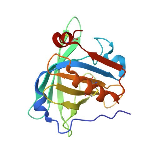
Reindeer beta-lactoglobulin crystal structure with pseudo-body-centred noncrystallographic symmetry.

Oksanen, E.Jaakola, V.P.Tolonen, T.Valkonen, K.Akerstrom, B.Kalkkinen, N.Virtanen, V.Goldman, A.

(2006) Acta Crystallogr D Biol Crystallogr 62: 1369-1374

  • DOI: https://doi.org/10.1107/S0907444906031519
  • Primary Citation Related Structures: 
    1YUP

  • PubMed Abstract: 

    Reindeer beta-lactoglobulin (betaLG) belongs to the lipocalin superfamily. Its DNA and protein sequences have been determined and showed that it had nine residue changes from bovine betaLG. Reindeer betaLG, the structure of which was finally determined at 2.1 A resolution in space group P1, crystallized in a unit cell that is both P2-like and P2(1)-like owing to the presence of an almost perfect (but noncrystallographic) body-centring vector. The non-body-centred data could only be observed using a very bright synchrotron beam and a novel refinement strategy was adopted to enable us to use the weak h + k + l = 2n + 1 reflections.


  • Organizational Affiliation
    • Institute of Biotechnology, University of Helsinki, Finland.

Macromolecule Content 

  • Total Structure Weight: 146.37 kDa 
  • Atom Count: 10,045 
  • Modeled Residue Count: 1,263 
  • Deposited Residue Count: 1,296 
  • Unique protein chains: 1

Macromolecules

Find similar proteins by:|  3D Structure
Entity ID: 1
MoleculeChains  Sequence LengthOrganismDetailsImage
beta-lactoglobulin
A, B, C, D, E
A, B, C, D, E, F, G, H
162Rangifer tarandusMutation(s): 0 
UniProt
Find proteins for P02754 (Bos taurus)
Go to UniProtKB:  P02754
Find proteins for Q00P86 (Rangifer tarandus tarandus)
Explore Q00P86 
Go to UniProtKB:  Q00P86
Entity Groups
Sequence Clusters30% Identity50% Identity70% Identity90% Identity95% Identity100% Identity
UniProt GroupQ00P86
Sequence Annotations
Expand
Reference Sequence

Experimental Data & Validation

Experimental Data

  • Method: X-RAY DIFFRACTION
  • Resolution: 2.10 Å
  • R-Value Free:  0.311 (Depositor), 0.304 (DCC) 
  • R-Value Work:  0.235 (Depositor), 0.236 (DCC) 
  • R-Value Observed: 0.239 (Depositor) 
Space Group: P 1
Unit Cell:
Length ( Å )Angle ( ˚ )
a = 52.24α = 90
b = 94.14β = 105.76
c = 64.15γ = 90
Software Package:
Software NamePurpose
REFMACrefinement
ProDCdata collection
XDSdata scaling
MOLREPphasing

Structure Validation

View Full Validation Report


Entry History 

Deposition Data

Revision History  (Full details and data files)

  • Version 1.0: 2006-02-14
    Type: Initial release
  • Version 1.1: 2008-04-30
    Changes: Version format compliance
  • Version 1.2: 2011-07-13
    Changes: Version format compliance
  • Version 1.3: 2017-10-11
    Changes: Refinement description
  • Version 1.4: 2023-10-25
    Changes: Data collection, Database references, Refinement description
  • Version 1.5: 2024-11-20
    Changes: Structure summary